 Back
BackChapter 1
Study Guide - Smart Notes
 Tailored notes based on your materials, expanded with key definitions, examples, and context.
Scope of Microbiology
Introduction
Microbiology is the study of microscopic organisms, including bacteria, viruses, fungi, protozoa, and algae. This field explores the structure, function, classification, and significance of microorganisms in health, disease, and the environment.
Achievements of Scientists in Early Microbiology
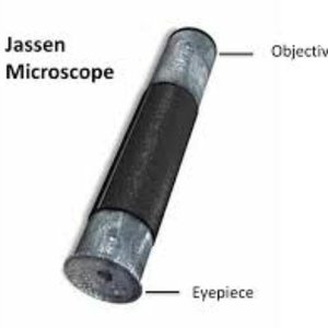
Zaccharias and Hans Janssen
Invention of the Compound Microscope: Dutch eyeglass makers who created the first compound microscope, consisting of a simple tube with lenses at each end. Magnification ranged from 3x to 9x depending on light.
Significance: Enabled the observation of objects too small for the naked eye, laying the foundation for microbiology.
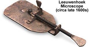
Antony van Leeuwenhoek- FIRST TO LOOK
Father of Microscopy: First to observe live bacteria and protozoans using a simple, single-lens microscope.
Discovery of Animalcules: Described microscopic life forms, which he called "animalcules." Did not connect them to disease.
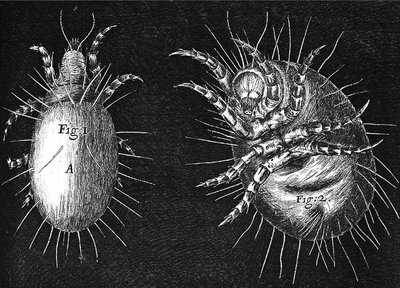
Robert Hooke
Improvements to the Compound Microscope: Enhanced the design and used it to observe various specimens, including plant cells and insects.
Publication: Authored Micrographia, illustrating his microscopic observations and confirming Leeuwenhoek’s findings.
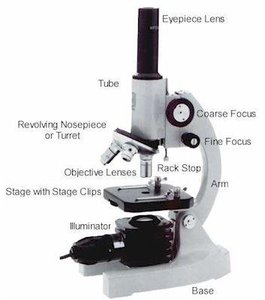
Types of Microscopes and Their Use
Light Microscopes
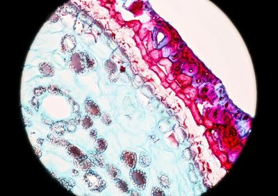
Light microscopes use visible light and optical lenses to magnify specimens. They are classified as simple (one lens) or compound (multiple lenses).
Ocular Lens: Located in the eyepiece.
Objective Lens: Located in the body of the microscope.
Total Magnification: Calculated as objective lens power × ocular lens power (e.g., 10x ocular × 4x objective = 40x).
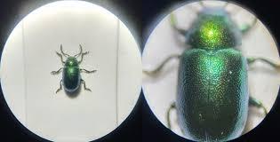
Stereomicroscope (Dissection Microscope)
Low Magnification: Used for viewing larger, three-dimensional objects, often during dissection.
Application: Provides a 3D view of specimens such as insects or plant parts.
Specialized Light Microscopy Techniques
Bright-Field: Produces a bright background; specimens often require staining.
Dark-Field: Illuminates specimen edges against a dark background; useful for observing motility and hard-to-stain organisms.
Phase-Contrast: Enhances contrast in transparent specimens by exploiting differences in refractive index.
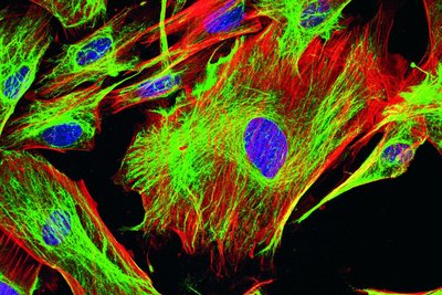
Fluorescence: Uses UV light to excite fluorescent dyes in specimens; widely used in diagnostics.
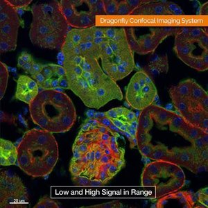
Confocal: Uses lasers to produce sharp, high-resolution images of thick specimens.
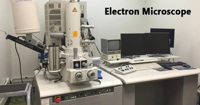
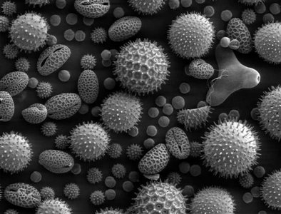
Electron Microscopes
Electron microscopes use electron beams and magnetic fields for much higher resolution than light microscopes. They require extensive sample preparation and are used for detailed cellular and molecular studies.
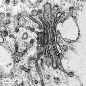
Transmission Electron Microscope (TEM): Electrons pass through ultra-thin specimens, producing 2D images with magnification up to 1,000,000x.
Scanning Electron Microscope (SEM): Scans the surface of specimens, producing detailed 3D images with magnification up to 100,000x.
Theory of Spontaneous Generation
Historical Experiments and Key Figures
Francesco Redi (1668): Demonstrated that maggots on meat came from fly eggs, not spontaneous generation.
John Needham (1745): Claimed spontaneous generation after observing microbial growth in boiled broth.
Lazzaro Spallanzani: Showed that boiling broth in sealed flasks prevented growth, suggesting microbes came from the air.
Louis Pasteur (1861): Used swan-neck flasks to show that microbes in the air, not spontaneous generation, caused growth in broth.
John Tyndall & Ferdinand Cohn: Provided evidence for heat-resistant bacteria and endospores.
Pasteurization: Developed by Pasteur, this process heats liquids to reduce microbial load and prevent spoilage, without sterilizing completely. THIS PROCCES DOES NOT KILL ALL MICROORGANISMS
Germ Theory of Disease and Its Significance
Development and Impact
Oliver Wendell Holmes & Ignaz Semmelweis: Demonstrated the importance of hand hygiene in preventing disease transmission.
Joseph Lister: Introduced aseptic techniques in surgery, reducing infections using carbolic acid.
Louis Pasteur & Robert Koch: Established that specific microbes cause specific diseases, forming the basis of the germ theory.
Edward Jenner: Developed the first vaccine (smallpox) using cowpox virus, founding immunology.
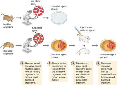
Koch’s Postulates
Criteria for Linking Microbes to Disease
The suspected causative agent must be absent from all healthy organisms but present in all diseased organisms.
The agent must be isolated from the diseased organism and grown in pure culture.
The cultured agent must cause the same disease when inoculated into a healthy, susceptible organism.
The same agent must be reisolated from the newly diseased organism.
Origin and Evolution of Microorganisms
Timeline and Evolutionary Relationships
Earth’s Formation: About 4.5 billion years ago; life began between 3.5 and 4 billion years ago.
First Life Forms: Prokaryotes were the earliest organisms, with eukaryotes evolving later (~2.2 billion years ago).
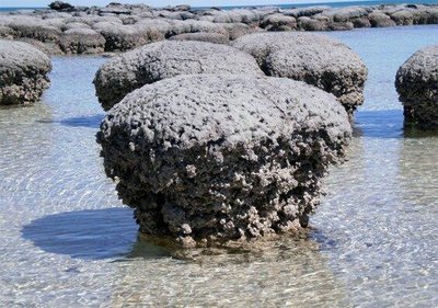
Stromatolites: Fossilized microbial mats, evidence of ancient microbial life.
Phylogeny: Evolutionary relationships are now determined using nucleic acid sequencing, classifying life into three domains: Bacteria, Archaea, and Eukarya.
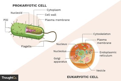
Differences Between Prokaryotes and Eukaryotes
Cellular Organization and Diversity
Prokaryotes: Single-celled organisms lacking a nucleus and membrane-bound organelles. Includes Bacteria and Archaea.
Bacteria: Found in diverse environments; some are pathogenic.
Archaea: Evolutionarily distinct from bacteria; often inhabit extreme environments.
Eukaryotes: Have a nucleus and membrane-bound organelles. Includes algae, fungi, and protozoans.
Viruses, Prions, Viroids: Non-cellular infectious agents; viruses consist of nucleic acid and protein, prions are misfolded proteins, and viroids are small RNA molecules infecting plants.
Role of Taxonomy
Classification and Nomenclature
Taxonomy: The science of organizing, classifying, and naming living organisms, introduced by Carolus Linnaeus.
Taxa: Non-overlapping groups based on similarities. Cats/DOGS
Classification: Assignment of organisms into taxa.
Nomenclature: Rules for naming organisms using a binomial system (Genus species).
Identification: Determining and recording traits of organisms.
Example: Homo sapiens (human), Apis mellifera (honeybee).
Second word lower case first word abreviated H.Sapiens
KING PHILLIP CAN ONLY FIND GREEN SOCKS- Classification
Microbial Transmission
Modes and Relationships
Biofilms: Communities of microbes within a polymeric matrix on surfaces.
Microbial Relationships:
Mutualism: Both organisms benefit.
Commensalism: One benefits, the other is unaffected.
Synergism: Both depend on each other to perform a function.
Parasitism: One benefits at the expense of the other.
Transmission Routes:
Waterborne: Through contaminated water; major cause of illness in developing countries.
Foodborne: Through contaminated food.
Airborne: Via aerosols from coughing, sneezing, or talking.
Zoonotic: Transmitted from animals to humans.
Uses of Microorganisms in Everyday Life
Applications and Benefits
Food Production: Microbes are essential in making vinegar, sauerkraut, pickles, yogurt, cheese, bread, and alcoholic beverages.
Water Treatment: Microbial analysis ensures safe drinking water.
Pharmaceuticals: Production of antibiotics (e.g., penicillin), hormones, and other drugs.
Agriculture: Microbes manage plant disease, soil fertility, and crop growth.
Bioremediation: Use of microbes to degrade toxic or hazardous substances.
Energy: Microbes convert organic material into alternative fuels (e.g., methane from landfills).
Forensics: Microbial analysis aids in solving crimes and tracing outbreaks.
