 Back
Selective and Differential Media in Microbiology: Principles and Applications
Study Guide - Smart Notes
 Tailored notes based on your materials, expanded with key definitions, examples, and context.
Selective and Differential Media
Overview of Culture Media
Culturing microbes in the laboratory requires specialized media that support the growth of specific organisms while inhibiting others. Selective, differential, and enrichment media are essential tools for isolating and identifying microbes in clinical and environmental microbiology.
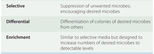
Type | Purpose |
|---|---|
Selective | Suppression of unwanted microbes; encouraging desired microbes |
Differential | Differentiation of colonies of desired microbes from others |
Enrichment | Similar to selective media but designed to increase numbers of desired microbes to detectable levels |

Definitions and Applications
Selective Media: Contains inhibitors (e.g., antibiotics, bile salts, high salt concentrations) that suppress the growth of unwanted microbes while allowing the target group to grow.
Differential Media: Contains substrates and indicators that allow visible distinction between different groups of microbes based on metabolic reactions (e.g., color changes due to pH indicators).
Enrichment Media: Similar to selective media but specifically formulated to enhance the growth of a particular microbe to detectable levels.
These media are widely used in clinical labs to isolate pathogens from normal flora and in environmental labs to detect contamination, such as coliforms in water.
Components of Selective and Differential Media
Key Components
Nutritional Components: Supply essential elements (C, N, P, etc.) and maintain appropriate pH for microbial growth. Often provided as digests or extracts (e.g., yeast extract, casein digest).
Inhibitors (Selective): Chemicals that prevent the growth of certain microbes (e.g., antibiotics, NaCl, bile salts).
Substrates (Differential): Compounds metabolized by microbes, allowing differentiation (e.g., lactose, mannitol).
Indicators (Differential): Chemicals that reveal metabolic changes (e.g., pH indicators like phenol red, neutral red, bromthymol blue; ferric ammonium citrate for sulfur reduction).
Examples of Selective and Differential Media
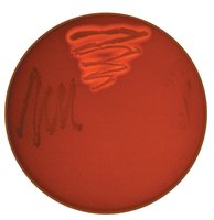
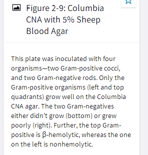
Columbia CNA with 5% Sheep Blood Agar
This medium is both selective and differential. It is used to isolate Gram-positive organisms and differentiate them based on hemolytic activity.
Selective: Colistin and nalidixic acid inhibit Gram-negative bacteria, allowing only Gram-positive organisms to grow.
Differential: Blood agar allows detection of hemolysis (lysis of red blood cells) by hemolysins.
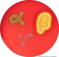
Types of Hemolysis:
Gamma (γ): No hemolysis; no color change around colonies.
Alpha (α): Partial hemolysis; greenish color around colonies.
Beta (β): Complete hemolysis; clear zone around colonies.
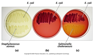
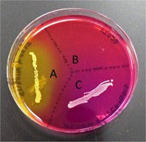
Mannitol Salt Agar (MSA)
MSA is used for the isolation and identification of Staphylococcus aureus from clinical specimens.
Selective: High salt concentration (7.5% NaCl) inhibits most bacteria except staphylococci and other halophiles.
Differential: Mannitol fermentation is detected by the pH indicator phenol red (yellow below pH 6.8 indicates fermentation).
Interpretation:
No growth: Not Staphylococcus.
Yellow media/colonies: Staphylococcus mannitol fermenter (likely S. aureus).
Pink media/colonies: Staphylococcus, non-mannitol fermenter (likely not pathogenic).
MacConkey Agar
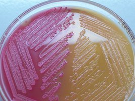
MacConkey agar is used to isolate and differentiate members of the family Enterobacteriaceae based on lactose fermentation.
Selective: Bile salts and crystal violet inhibit Gram-positive bacteria, allowing only Gram-negative bacteria to grow.
Differential: Lactose fermentation is detected by the pH indicator neutral red (pink below pH 6.8).
Interpretation:
No growth: Gram-positive organism.
Pink colonies: Gram-negative, lactose fermenter (e.g., Escherichia coli).
Yellow/colorless colonies: Gram-negative, non-lactose fermenter (e.g., Salmonella).
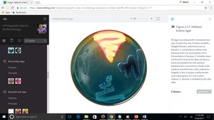
Hektoen Enteric (HE) Agar
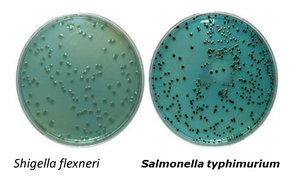
HE agar is used to isolate and differentiate Salmonella and Shigella from other Gram-negative enterics.
Selective: Bile salts inhibit Gram-positive bacteria, allowing only Gram-negative bacteria to grow.
Differential:
Sugar fermentation: Lactose, sucrose, and salicin fermentation detected by bromthymol blue and acid fuchsin (yellow or pink colonies indicate fermentation).
Sulfur reduction: Sodium thiosulfate (substrate) and ferric ammonium citrate (indicator) detect H2S production (black precipitate indicates sulfur reduction).
Interpretation:
No growth: Gram-positive organism.
Yellow, orange, or pink colonies: Gram-negative, ferments at least one sugar, does not reduce sulfur.
Blue or green colonies: Gram-negative, does not ferment sugars, does not reduce sulfur (possible Salmonella or Shigella).
Black colonies: Gram-negative, does not ferment sugars, reduces sulfur (possible Salmonella).
Fermentation and Differential Media
Principles of Fermentation in Microbial Identification
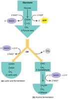
Many differential media rely on the ability of microbes to ferment specific substrates, producing acidic byproducts that lower the pH of the medium. pH indicators are used to detect these changes, allowing for the identification of fermenters versus non-fermenters.
Fermentation: Metabolic process that converts sugars to acids, gases, or alcohol in the absence of oxygen.
pH Indicators: Chemicals that change color in response to pH shifts, revealing fermentation activity.
Summary Table: Common Selective and Differential Media
Medium | Selective For | Differential For | Key Indicator(s) | Interpretation |
|---|---|---|---|---|
Columbia CNA with Blood | Gram-positive bacteria | Hemolysis | Blood (hemolysis) | α, β, γ hemolysis |
Mannitol Salt Agar | Staphylococci | Mannitol fermentation | Phenol red | Yellow = fermenter |
MacConkey Agar | Gram-negative rods | Lactose fermentation | Neutral red | Pink = fermenter |
Hektoen Enteric Agar | Gram-negative enterics | Sugar fermentation, sulfur reduction | Bromthymol blue, acid fuchsin, ferric ammonium citrate | Yellow/pink = fermenter; black = sulfur reducer |
Additional info: Selective and differential media are foundational tools in microbiology for isolating and identifying pathogens, monitoring environmental contamination, and studying microbial physiology. Understanding the principles behind these media and interpreting their results is essential for laboratory diagnostics and research.
