 Back
Simple Stain and Smear Preparation: Principles and Laboratory Techniques
Study Guide - Smart Notes
 Tailored notes based on your materials, expanded with key definitions, examples, and context.
Simple Stain and Smear Preparation
Principles of Staining
Staining is a fundamental technique in microbiology used to enhance the visibility of microorganisms under a microscope. By increasing contrast between cells and their background, stains allow for easier observation of cell morphology and arrangement.
Simple stains use a single dye and reveal basic cell features such as shape, size, and arrangement.
Differential stains use multiple dyes to distinguish between different types of cells or cell structures, often based on cell wall composition.
Chromophores are ions in staining solutions that impart color. Their chemical nature determines their interaction with cells.
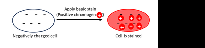
Basic dyes (e.g., methylene blue, crystal violet, safranin) are cationic and bind to negatively charged bacterial cells, staining the cells directly.
Acidic dyes (e.g., nigrosine, Congo red) are anionic and are repelled by bacterial cells, staining the background instead.
Example: Methylene blue is a basic dye commonly used for simple staining.
Smear Preparation and Fixation
Preparing a smear is an essential step before staining. A smear is a thin layer of specimen dried and fixed onto a glass slide. Fixation ensures the sample adheres to the slide and kills most cells, making them safer to handle.
Heat fixation is the most common method, performed by passing the slide through a Bunsen burner flame.
Chemical fixation uses chemical agents to fix cells, but is less commonly used in basic laboratory procedures.
Proper smear preparation is crucial for effective staining and observation.
Example: Smears can be prepared from oral samples, liquid cultures, or solid cultures.
Laboratory Procedure: Simple Stain
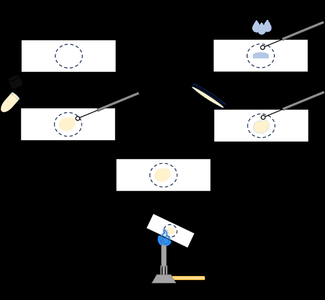
The simple staining procedure involves preparing a smear, fixing it, applying a stain, rinsing, drying, and observing under a microscope.
Prepare the smear (from oral, liquid, or solid sample).
Allow the smear to air dry until it appears cloudy.
Heat fix the sample by passing the slide through a flame 2-3 times.
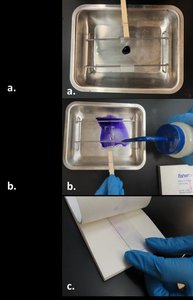
Place the slide over a staining tray and cover the smear with methylene blue for 1 minute.
Rinse the slide gently with water until excess dye is removed.
Blot dry using bibulous paper, avoiding rubbing or dragging.
Observe the stained smear under a compound light microscope, up to oil immersion.
Interpretation and Observations
After staining, students should record observations regarding specimen morphology, arrangement, and total magnification used. Simple stains allow identification of cell shape (e.g., cocci, bacilli), arrangement (e.g., clusters, chains), and size.
Cell morphology: Refers to the shape of the cells (e.g., round, rod-shaped).
Cell arrangement: Describes how cells are grouped (e.g., pairs, chains, clusters).
Magnification: Oil immersion (1000x) is often required to observe bacterial cells clearly.
Example: Oral smears may show a variety of bacterial morphologies and arrangements, as well as larger eukaryotic cheek cells.
Key Questions and Concepts
True or False: Simple staining requires only one dye and a fixed smear; it is not performed on live samples.
Purpose of heat-fixing: To adhere cells to the slide and kill them for safer handling.
Alternative dyes: Crystal violet and safranin can be used for simple staining.
Simple stain reveals: Cell shape, size, and arrangement (not cell wall composition).
Features of basic dye: Contains a chromophore, has a positive charge, stains the cell (not the background).
Heat fixation effects: May alter cell morphology or cause cell shrinkage.
Poor staining outcomes: May result from insufficient sample or improper fixation.
Summary Table: Simple vs Differential Stains
Stain Type | Dyes Used | Information Revealed | Examples |
|---|---|---|---|
Simple Stain | One | Cell shape, size, arrangement | Methylene blue, crystal violet |
Differential Stain | Two or more | Cell wall composition, cell type | Gram stain, acid-fast stain |
Additional info:
Proper smear thickness is important; too thick or too thin smears can hinder observation.
Oil immersion increases resolution for observing small bacterial cells.
