 Back
BackSkin and Throat Microbiota: Laboratory Study Guide
Study Guide - Smart Notes
 Tailored notes based on your materials, expanded with key definitions, examples, and context.
Skin Flora
Introduction to Skin Microbiota
The human skin is colonized by a diverse community of microorganisms, collectively known as the skin microbiota. Approximately 2 m2 of skin surface harbors around 1,000 different bacterial species from 19 phyla, with an estimated total of 1012 bacteria. Most reside on the superficial epidermis and upper hair follicles. The majority of these microbes are non-pathogenic and provide benefits such as occupying host cell receptors, thereby preventing colonization by pathogenic organisms.
Resident microbiota: Permanent inhabitants of the skin, generally harmless or beneficial.
Opportunistic pathogens: Resident microbes that can cause disease if the skin barrier is breached (e.g., cellulitis, septicemia).
Transmission: Direct contact (with people or fomites) is a key route for spreading microbes.
Handwashing and Microbial Transmission
Handwashing is a critical method for reducing the transmission of potentially harmful microbes, especially in healthcare and food service settings. Laboratory experiments culturing the tactile surfaces of fingers before and after washing demonstrate the effectiveness of hand hygiene.
Prediction: One might expect fewer bacteria after washing, but experiments often show an increase in colony numbers post-washing, though with reduced diversity.
Explanation: Washing removes transient, detergent-susceptible bacteria, leaving behind tightly adhered resident microbiota.
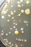
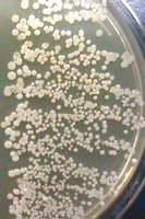
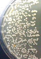
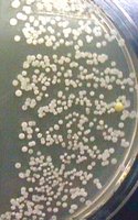
Example: After 2, 4, and 6 minutes of handwashing, the number of colonies increases, but the types of colonies decrease, indicating removal of transient flora and persistence of resident flora.
Staphylococci on the Skin
Members of the genus Staphylococcus are common skin and mucous membrane microbiota. While most are harmless, all can be opportunistic pathogens, and Staphylococcus aureus is a significant pathogen causing various infections and food poisoning. Differentiation between S. aureus and other staphylococci is crucial in clinical microbiology.
Blood agar: Used to assess hemolysis; β-hemolysis is associated with pathogenic strains.
Mannitol Salt Agar (MSA): Selective-differential medium for Staphylococcus spp.
Mannitol Salt Agar (MSA)
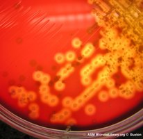
MSA contains 7.5% NaCl (selective agent) and mannitol (differential agent). The high salt concentration inhibits most bacteria except Staphylococcus spp. S. aureus ferments mannitol, producing acid that turns the phenol red indicator yellow, while most other staphylococci do not ferment mannitol.
Selection: High NaCl inhibits non-staphylococci.
Differentiation: S. aureus ferments mannitol (yellow), others do not (red).
Coagulase Test
The coagulase test distinguishes S. aureus (coagulase-positive) from other staphylococci (coagulase-negative). Coagulase exists in two forms: free and bound (clumping factor).
Tube Test: Detects both free and bound coagulase using rabbit plasma; clotting indicates a positive result.
Slide Test: Detects only bound coagulase; agglutination within 1–2 minutes indicates positivity.
Biochemical pathway:
Free coagulase: Activates prothrombin to thrombin, which converts fibrinogen to fibrin (clot).
Bound coagulase: Directly activates fibrinogen.
Latex Agglutination Test
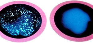
The BACTI™ STAPH Latex Slide Agglutination Test uses latex beads coated with proteins that react with bound coagulase and protein A, both present in S. aureus cell walls. Agglutination indicates a positive result for S. aureus.
Throat Flora
Introduction to Throat Microbiota
The upper respiratory tract (URT) harbors a diverse microbiota, including organisms found on the skin and many others. Most are non-pathogenic and beneficial, but under certain conditions, resident microbes can become opportunistic pathogens, causing abscesses or septicemia if mucous membranes are breached. Coughing and sneezing facilitate microbe transmission. Streptococcus pyogenes and Streptococcus pneumoniae are particularly important in human disease.
Streptococci: Identification and Classification
Streptococci are initially cultured on blood agar to assess hemolysis, which is key for identification. They are also classified into serological groups (A, B, C, D, etc.) based on cell wall polysaccharides. Sensitivity to antibiotics (e.g., bacitracin, optochin) further differentiates species.
Group A Streptococcus (S. pyogenes): Bacitracin-sensitive.
S. pneumoniae: Optochin-sensitive.
Throat Swab and Hemolysis
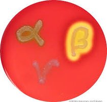
Throat swabs are commonly used to sample the palatine tonsils for URT infections. Culturing on sheep blood agar allows for the evaluation of hemolysis:
α-hemolysis: Partial hemolysis, green halo (viridans streptococci).
β-hemolysis: Complete hemolysis, clear halo (S. pyogenes).
γ-hemolysis: No hemolysis.
Catalase Test: Differentiates staphylococci (catalase-positive) from streptococci (catalase-negative). Note: Never perform directly on blood agar to avoid false positives.
Streptolysins
Streptolysin S and Streptolysin O are β-hemolysins produced by S. pyogenes:
Streptolysin S: Oxygen-stable, detected on the surface of blood agar.
Streptolysin O: Oxygen-labile, detected by stabbing the agar (less oxygen below the surface).
Stabbing the blood agar plate enhances detection of oxygen-sensitive hemolysins, resulting in sharper β-hemolysis zones in the stab compared to the surface.
Rapid Group A Streptococcus Antigen Test
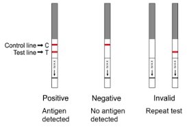
This is a rapid immunochromatographic assay for detecting group A streptococcal antigen directly from throat swabs. Results are interpreted as follows:
Result | Interpretation |
|---|---|
Two lines (control + test) | Positive: Antigen detected |
One line (control only) | Negative: No antigen detected |
No control line | Invalid: Repeat test |
Summary Table: Key Laboratory Media and Tests
Test/Medium | Purpose | Key Result |
|---|---|---|
Blood Agar | Hemolysis pattern | α, β, or γ hemolysis |
Mannitol Salt Agar | Selection/Differentiation of Staphylococci | Yellow for S. aureus, red for others |
Coagulase Test | Differentiation of S. aureus | Clotting/agglutination = positive |
Latex Agglutination | Rapid S. aureus ID | Agglutination = positive |
Rapid Strep Test | Group A Strep detection | Two lines = positive |
