 Back
Structural Stains in Microbiology: Capsules, Endospores, and Flagella
Study Guide - Smart Notes
 Tailored notes based on your materials, expanded with key definitions, examples, and context.
Structural Stains in Microbiology
Introduction to Structural Stains
Structural stains are specialized microbiological techniques used to visualize specific bacterial structures that are otherwise difficult to observe with standard staining methods. These include the capsule, endospore, and flagella, each of which plays a critical role in bacterial physiology and pathogenicity.
Capsule Stain: Reveals the protective polysaccharide or polypeptide layer surrounding some bacteria.
Endospore Stain: Highlights dormant, highly resistant structures formed by certain genera.
Flagella Stain: Visualizes the motility appendages of bacteria.
Capsules
Structure and Function of Capsules
Capsules are extracellular layers composed mainly of polysaccharides (sometimes polypeptides) that surround the cell wall of certain bacteria.
Protection: Capsules protect bacteria from phagocytosis by host immune cells, enhancing pathogenicity.
Composition: Most capsules are made of polysaccharides, though some are polypeptide-based.
Visualization: Capsules are visualized using negative staining techniques, where the capsule remains unstained and appears as a clear halo around the stained cell and background.
Example: Klebsiella pneumoniae produces a prominent capsule, aiding its ability to cause pneumonia. 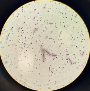
Endospores
Structure and Function of Endospores
Endospores are highly resistant, dormant structures formed by certain bacteria (notably Bacillus and Clostridium) in response to environmental stress.
Resistance: Endospores withstand extreme heat, radiation, and chemicals.
Formation: Endospore formation (sporulation) is not a reproductive process but a survival mechanism.
Staining: Endospores require heat to allow stains to penetrate. After staining, endospores retain the primary stain, while vegetative cells are counterstained.
Positions: Endospores can be central, terminal, or subterminal within the cell.
Example: Bacillus subtilis forms endospores that allow it to survive harsh conditions. 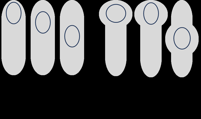
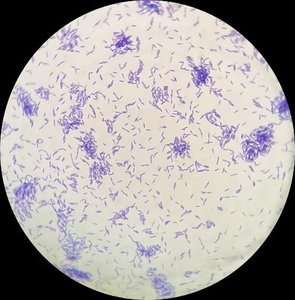
Endospore Staining Procedure
Heat is used to drive the primary stain (usually malachite green) into the endospore.
Vegetative cells are decolorized and counterstained with safranin, appearing pink/red.
Endospores retain the green color.
Formula: Table: Endospore Positions
Position | Description |
|---|---|
Central | Endospore located in the middle of the cell |
Terminal | Endospore at the end of the cell |
Subterminal | Endospore between the center and end |
Swollen | Endospore causes cell to swell |
Flagella
Structure and Function of Flagella
Flagella are long, thin, proteinaceous appendages that provide motility to bacteria.
Motility: Flagella enable bacteria to move toward favorable environments or away from harmful stimuli (chemotaxis).
Arrangement: Flagella can be arranged in several ways: polar (at one or both ends), peritrichous (all around the cell), amphitrichous (at both ends), etc.
Staining: Flagella are too thin to be seen with light microscopy unless stained and coated with a mordant to increase their diameter.
Example: Escherichia coli exhibits peritrichous flagella, contributing to its motility. 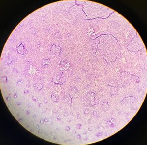
Flagella Arrangements
Monotrichous: Single flagellum at one end
Amphitrichous: Single flagellum at both ends
Lophotrichous: Tuft of flagella at one or both ends
Peritrichous: Flagella distributed over the entire cell surface
Pathogenicity and Structural Features
Role in Pathogenicity
Capsules: Enhance pathogenicity by preventing phagocytosis, allowing bacteria to evade the immune system.
Endospores: Allow survival in hostile environments, contributing to persistence and transmission of disease.
Flagella: Aid in motility, helping bacteria reach target tissues and colonize hosts.
Summary Table: Structural Stains
Structure | Staining Method | Function | Pathogenicity |
|---|---|---|---|
Capsule | Negative stain + basic dye | Protection from phagocytosis | Evades immune response |
Endospore | Heat + malachite green, counterstain safranin | Survival in harsh conditions | Persistence, transmission |
Flagella | Mordant + stain | Motility | Colonization, invasion |
Lab Report and Questions
Key Points for Lab Report
Record observations for each structure (morphology, position, arrangement, magnification).
Compare endospore samples and explain differences (e.g., age, environmental conditions).
Interpret staining results: endospores appear green, cells appear red/purple.
Sample Questions and Answers
Endospores serve as reproductive structures: False. They are survival structures.
Capsule staining technique incorporates negative staining: True.
Flagella staining requires a mordant: True.
Capsules are: Extracellular structures.
Process of endospore formation: Sporulation.
Endospores are dormant structures: True.
Flagella contribute to pathogenicity: By enabling motility, bacteria can invade host tissues and escape immune responses.
Capsules contribute to pathogenicity: By preventing phagocytosis, capsules allow bacteria to survive and cause disease.
Additional info: Academic context was added to clarify staining methods, structural functions, and pathogenicity roles.
