 Back
BackStructural Stains in Microbiology: Endospores, Capsules, and Flagella
Study Guide - Smart Notes
 Tailored notes based on your materials, expanded with key definitions, examples, and context.
Structural Stains in Microbiology
Introduction to Structural Stains
Structural stains are specialized microbiological techniques used to visualize and study specific bacterial structures such as endospores, capsules, and flagella. These structures are often too small or lack sufficient contrast to be seen with standard light microscopy, so special dyes and reagents are employed to highlight them. Understanding these stains is crucial for bacterial identification, classification, and understanding pathogenic mechanisms.
Endospores
Definition and Biological Role
Endospores are highly resistant, dormant structures formed by certain bacteria, primarily in the orders Bacillales and Clostridiales.
They are not reproductive structures but serve as survival forms, allowing bacteria to withstand extreme conditions such as heat, desiccation, radiation, and chemicals.
Endospores can remain viable for thousands or even millions of years, reactivating when environmental conditions become favorable.
Clinical relevance: Endospore-forming bacteria (e.g., Bacillus anthracis, Clostridium tetani, Clostridium difficile) are difficult to eradicate due to their resistance to disinfectants and persistence in the environment.
Structure of Endospores
Exosporium: Thin outer covering for protection.
Coats: Protein shields that provide chemical and enzymatic resistance.
Cortex: Thick peptidoglycan layer that keeps the core dry, contributing to heat resistance.
Core wall: Becomes the future cell wall upon germination.
Core: Contains DNA, ribosomes, and essential enzymes.
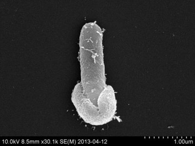
Endospore Formation (Sporulation)
Sporulation is the process by which certain Gram-positive bacteria form endospores in response to unfavorable conditions such as nutrient deprivation, heat, or desiccation.
Vegetative Cycle: Under nutrient-rich conditions, bacteria grow and divide by binary fission.
Asymmetric Cell Division: Upon stress, the cell divides unequally, forming a larger mother cell and a smaller forespore.
Engulfment: The mother cell engulfs the forespore, providing it with a second membrane.
Spore Coat and Cortex Formation: Protective layers (inner and outer membranes, cortex, spore coat) form around the forespore.
Late Sporulation: The spore matures, accumulating dipicolinic acid and calcium, which stabilize DNA and proteins.
Mother Cell Lysis: The mother cell lyses, releasing the mature spore into the environment.
Germination (Return to Vegetative State)
Activation: Triggered by environmental signals such as nutrients.
Germination Proper: The spore cortex is degraded, water uptake increases, and metabolism resumes.
Outgrowth: The spore transforms into an actively dividing vegetative cell.
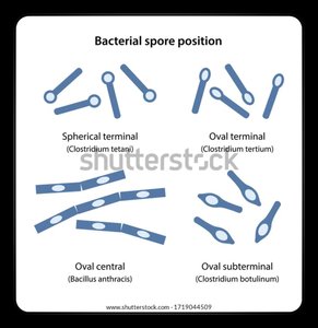
Types of Endospores
Endospores can be classified based on their position within the bacterial cell:
Spherical terminal (e.g., Clostridium tetani)
Oval terminal (e.g., Clostridium tertium)
Oval central (e.g., Bacillus anthracis)
Oval subterminal (e.g., Clostridium botulinum)
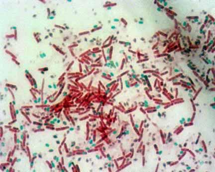
Endospore Staining
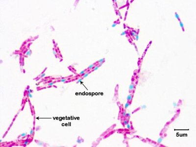
Endospores are impermeable to most stains, so heat is used to drive the stain into the spore. The Schaeffer-Fulton method is commonly used:
Primary stain: Malachite green (applied with heat)
Decolorization: Water (removes stain from vegetative cells, not endospores)
Counterstain: Safranin (stains vegetative cells red/pink)
Results: Endospores appear green; vegetative cells appear red/pink.
Examples and Longevity
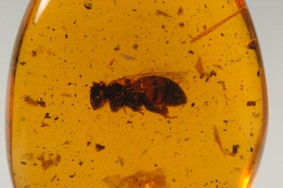
Viable endospores have been isolated from ancient sources, such as the GI tract of a bee in amber (25–40 million years old) and salt crystals (over 250 million years old).
Flagella
Structure and Function
Flagella are thin, proteinaceous appendages that provide motility to many bacteria.
They originate in the cytoplasm and project through the cell wall.
Flagella are too thin to be seen with a light microscope without special staining.
Types of motility: Flagellar (swimming), gliding (e.g., myxobacteria), and axial filaments (e.g., spirochetes).
Types of Flagellar Arrangement
Monotrichous: Single flagellum at one pole (e.g., Pseudomonas aeruginosa).
Lophotrichous: Tuft of flagella at one pole.
Amphitrichous: Flagella at both poles.
Peritrichous: Flagella distributed all over the cell (e.g., Escherichia coli, Proteus spp.).
Amphilophotrichous: Tuft of flagella at both ends.

Flagella Staining
Flagella are visualized using a mordant (e.g., tannic acid, potassium alum) to coat and thicken them, followed by a basic dye (e.g., carbolfuchsin or crystal violet).
This process allows the number and arrangement of flagella to be observed, aiding in bacterial identification.
Procedure:
Prepare a bacterial smear without harsh heat fixing.
Apply mordant to coat flagella.
Add basic dye to stain the flagella.
Rinse and observe under oil immersion.
Result: Flagella appear as thin, stained filaments extending from the cell body.
Capsules
Structure and Function
Capsules are thick, gelatinous outer layers composed of polysaccharides or polypeptides.
They protect bacteria from environmental hazards and host immune defenses, aid in adherence to surfaces, and contribute to biofilm formation.
Capsules are important virulence factors, preventing phagocytosis and enhancing pathogenicity (e.g., Streptococcus pneumoniae, Klebsiella pneumoniae).
Capsule Staining
Capsules do not readily take up most dyes due to their non-ionic, water-soluble nature.
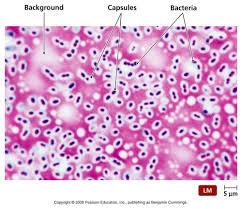
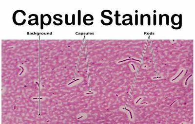
Negative staining is used: an acidic dye (e.g., India ink or nigrosin) stains the background, while a basic dye (e.g., crystal violet or safranin) stains the bacterial cell.
The capsule appears as a clear halo around the colored cell against a dark background.
Procedure:
Place a drop of India ink on a slide and mix with bacterial suspension.
Spread gently to create a thin smear.
Add a counterstain to color the bacterial cells.
Observe under oil immersion.
Result: Background is dark, bacterial cell is colored, and capsule is a clear halo.
Comparison of Structural Stains
Feature | Endospore Stain | Capsule Stain | Flagella Stain |
|---|---|---|---|
Purpose | Detect bacterial endospores (highly resistant dormant structures) | Visualize capsules (protective outer layers) | Visualize flagella (motility structures) |
Principle | Heat + malachite green to force dye into spore coat; safranin counterstain colors vegetative cells | Negative staining (India ink/nigrosin for background) + basic stain for cells; capsule = clear halo | Mordant (tannic acid/potassium alum) to coat & thicken, then stain with carbolfuchsin/crystal violet |
Procedure (simplified) | 1. Malachite green + heat 2. Rinse 3. Safranin counterstain | 1. India ink + bacteria 2. Counterstain with crystal violet/safranin | 1. Mordant 2. Basic dye 3. Observe under oil immersion |
Result | Spores = green, vegetative cells = red/pink | Capsule = clear halo, cell colored, background dark | Flagella visible as stained filaments; arrangement observed |
Examples of Organisms | Bacillus anthracis, Clostridium tetani, Clostridium difficile | Klebsiella pneumoniae, Streptococcus pneumoniae | Escherichia coli (peritrichous), Pseudomonas aeruginosa (monotrichous), Proteus spp. (peritrichous) |
Clinical Relevance | Persistence & resistance; important in sterilization and infection control | Virulence factor; prevents phagocytosis, key in meningitis & pneumonia | Motility & virulence; aids in UTI spread, species identification |
