 Back
Backlecture 12
Study Guide - Smart Notes
 Tailored notes based on your materials, expanded with key definitions, examples, and context.
Prokaryotic Pathogens: Bacterial Strategies for Survival and Pathogenesis
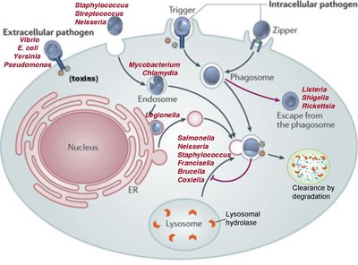
Extracellular vs. Intracellular Bacterial Pathogens
Bacteria can be classified based on their location relative to host cells during infection. This distinction is critical for understanding their pathogenic mechanisms and how the immune system responds to them.
Extracellular pathogens reside outside or on the surface of host cells, often producing toxins and enzymes to combat host defenses.
Intracellular pathogens invade and survive within host cells, evading immune detection and destruction.
Examples of extracellular pathogens: Staphylococcus aureus, Propionibacterium acnes, Helicobacter pylori, Pseudomonas aeruginosa, Vibrio cholerae.
Examples of intracellular pathogens: Listeria monocytogenes, Brucella abortus, Salmonella enterica, Shigella flexneri, Mycobacterium tuberculosis, Coxiella, Chlamydia.
Intracellular pathogens can evade immune responses by hiding within host cells, while extracellular pathogens rely on secreted factors to survive.
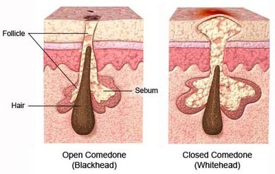
Acne: Skin Infection with Normal Microbiota
Acne is a common skin condition caused by the colonization of sebaceous glands by normal skin microbiota, particularly Propionibacterium acnes and Staphylococcus epidermidis.
Open comedone (blackhead): Occurs when dried sebum and dead skin cells plug the follicle, exposed to air and oxidized.
Closed comedone (whitehead): Formed when the plug is covered by skin, trapping sebum and creating an anaerobic environment.
The anaerobic environment promotes bacterial growth, leading to inflammation and infection.
Antibiotics and benzoyl peroxide are effective treatments.
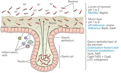
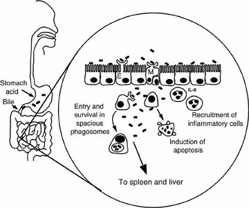
Gastritis, Ulcers, and Cancer: Helicobacter pylori Infection
Helicobacter pylori is a helical, motile Gram-negative bacterium adapted to the acidic environment of the stomach. It is associated with gastritis, peptic ulcers, and stomach cancer.
Uses flagella to penetrate the mucus lining and reach epithelial cells.
Produces urease, which converts urea to CO2 and ammonia, neutralizing stomach acid.
Colonization factors and immune modulators include adhesins and toxins.
Tissue Penetration and Dissemination
Spreading Factors and Enzymes
Pathogenic bacteria produce enzymes known as spreading factors to facilitate movement through host tissues and dissemination from the site of infection.
DNases: Degrade DNA in pus, reducing viscosity and aiding spread.
Hyaluronidases: Degrade hyaluronic acid in connective tissue, breaking down the extracellular matrix.
Collagenases and elastases: Proteases that degrade matrix proteins, aiding tissue invasion.
Plasminogen activator-like proteases: Dissolve fibrin clots, allowing escape from blood clots.
Staphylokinase: Produced by Staphylococcus aureus, activates plasminogen to plasmin, dissolving clots.
Exfoliative toxins: Cause scalded skin syndrome by blistering and peeling skin.
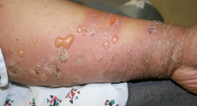
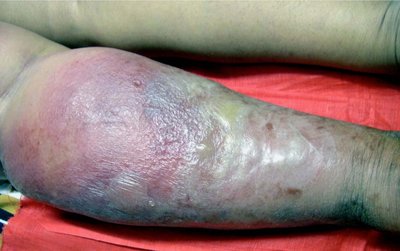
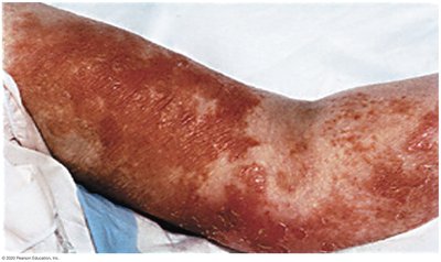
Clinical Manifestations of Extracellular Pathogens
Impetigo: Superficial skin infection with blisters and crusting.
Scalded skin syndrome: Blistering and peeling of skin, often in children.
Toxic shock syndrome: Systemic illness caused by toxin-producing strains.
Necrotizing fasciitis: Rapidly spreading infection causing tissue death.


Survival in the Host: Nutrient Acquisition
Enzymatic Breakdown of Host Macromolecules
Bacteria secrete enzymes to break down host macromolecules, releasing nutrients for their growth and survival.
Glycosidases: Hydrolyze sugars and polysaccharides (e.g., neuraminidases, sialidases, hyaluronidases).
Proteases: Degrade proteins, including matrix proteins and immunoglobulins.
Nucleases: Degrade DNA and RNA in pus.
Phospholipases: Cleave membrane lipids, lysing host cells.
Toxins: Disrupt host cells, releasing cytosolic contents.
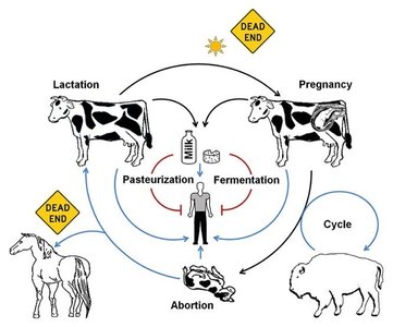
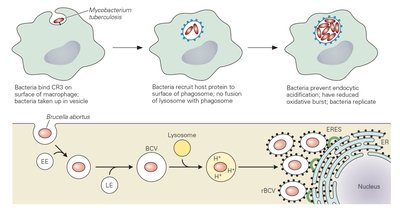
Example: Brucella abortus and Erythritol
Brucella abortus causes brucellosis, a zoonotic disease affecting domestic animals and humans. It grows rapidly in the placenta due to high erythritol concentrations.
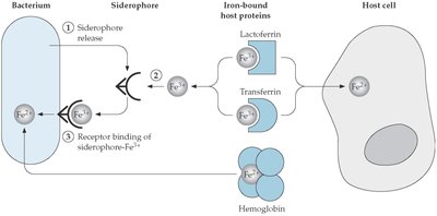
Survival in the Host: Iron Acquisition
Iron Limitation and Bacterial Strategies
Iron is essential for bacterial growth but is tightly sequestered in the host. Bacteria have evolved mechanisms to acquire iron from host proteins.
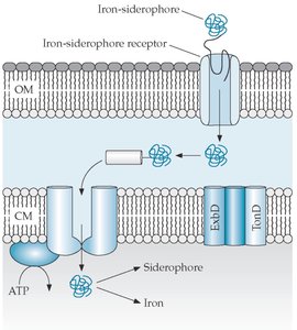
Siderophores: High-affinity iron-chelating molecules secreted by bacteria.
Receptors: Bacterial surface proteins bind host iron carriers (transferrin, lactoferrin, ferritin, heme).
Proteases: Degrade host proteins to release iron.
Alternative strategies: Some bacteria (e.g., Borrelia burgdorferi) use Mn2+ instead of Fe2+.
Cytolytic toxins: Lyse host cells to release iron.
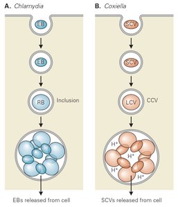
Intracellular Bacterial Pathogens: Survival Strategies
Obligate Intracellular Bacteria: Two Forms
Obligate intracellular bacteria, such as Chlamydia and Coxiella, have distinct forms for infection and replication.
Elementary Body (EB): Infectious, metabolically inactive form.
Reticulate Body (RB): Noninfectious, metabolically active, replicative form.
EBs are released from cells after conversion from RBs.
Specialized Compartments and Phagosome Escape
Intracellular pathogens survive by manipulating host cell compartments, preventing lysosomal degradation, or escaping into the cytosol.
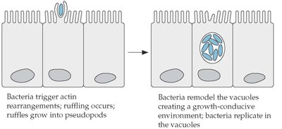
Salmonella enterica: Survives in specialized vacuoles within macrophages, preventing phagolysosomal fusion.
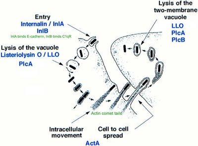
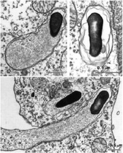
Listeria monocytogenes: Escapes the phagosome using pore-forming toxins, spreads cell-to-cell via actin-based motility.
Shigella flexneri: Uses pore-forming toxins to escape phagosomes.
Summary Table: Extracellular vs. Intracellular Pathogens
Pathogen Type | Location | Key Mechanisms | Examples |
|---|---|---|---|
Extracellular | Outside host cells | Toxins, spreading enzymes, immune evasion | Staphylococcus aureus, Propionibacterium acnes, Helicobacter pylori |
Intracellular | Inside host cells | Phagosome escape, specialized vacuoles, immune evasion | Listeria monocytogenes, Salmonella enterica, Chlamydia, Coxiella |
Key Terms and Concepts
Fimbriae: Surface structures for bacterial adhesion to host cells.
Phagosome: Intracellular vesicle containing engulfed pathogens.
Siderophores: Molecules secreted by bacteria to bind and transport iron.
Actin-based motility: Mechanism for intracellular movement and cell-to-cell spread.
Spreading factors: Enzymes that facilitate tissue invasion and dissemination.
Equations and Formulas
Iron chelation by siderophores can be represented as:
Urease reaction in Helicobacter pylori:
Additional info:
Some content was expanded for clarity and completeness, including definitions and examples of pathogenic mechanisms.
Images were included only when directly relevant to the explanation of the adjacent paragraph.
