 Back
BackStudy Notes: Chapter 13
Study Guide - Smart Notes
 Tailored notes based on your materials, expanded with key definitions, examples, and context.
Characterizing and Classifying Viruses, Viroids, and Prions
Characteristics of Viruses
Viruses are miniscule, acellular infectious agents that possess either DNA or RNA as their genetic material. They are responsible for numerous infections across humans, animals, plants, and bacteria, and cause many diseases in the industrialized world.
Viruses cannot carry out metabolic pathways independently, do not grow or respond to their environment, and cannot reproduce without a host cell.
They recruit the host cell's metabolic pathways to replicate.
Viruses lack cytoplasmic membrane, cytosol, and organelles (with rare exceptions).
They exist in two states: extracellular (virion) and intracellular (nucleic acid only).
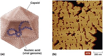
Extracellular State: Virion
The virion consists of a protein coat (capsid) surrounding the nucleic acid.
The combination of nucleic acid and capsid is called the nucleocapsid.
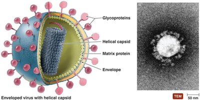
Some virions possess a phospholipid envelope.
The outermost layer provides protection and recognition sites for host cells.
Intracellular State
Once inside the host cell, the capsid is removed, and the virus exists as nucleic acid.
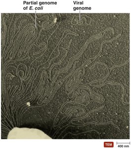
Genetic Material of Viruses
Viruses display greater diversity in their genomes compared to cells, which is a primary basis for their classification.
Viral genomes may be DNA or RNA, but never both.
They can be double-stranded (dsDNA, dsRNA) or single-stranded (ssDNA, ssRNA).
Genomes may be linear and segmented or single and circular.
Viral genomes are much smaller than those of cells.
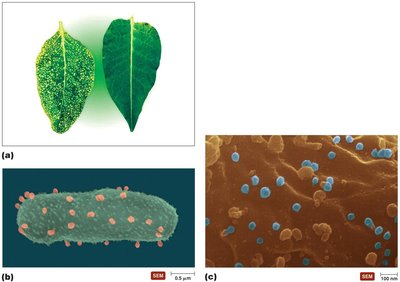
Hosts of Viruses
Viruses are highly specific to their hosts, often infecting only particular cells within a host due to the affinity of viral surface proteins for complementary host cell proteins.
Some viruses are generalists, infecting many kinds of cells in various hosts.
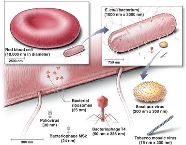
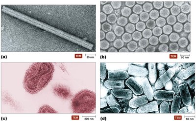
Sizes of Virions
Virions vary greatly in size, typically much smaller than most cells and organelles.
Capsid Morphology
The capsid is a protein coat that protects viral nucleic acid and facilitates attachment to host cells.
Capsids are composed of protein subunits called capsomeres.
Capsomeres may consist of one or several types of proteins.
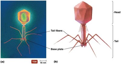
Shapes of Virions
Virions can be helical, polyhedral, or complex in shape.
Complex Shape of Bacteriophage T4
Bacteriophages often have a complex structure with a head, tail, tail fibers, and base plate.
The Viral Envelope
Some viruses acquire an envelope from the host cell during replication or release.
The envelope is a portion of the host's membrane system, composed of a phospholipid bilayer and proteins.
Virally coded glycoproteins (spikes) are often present and play a role in host recognition.
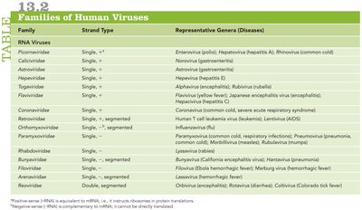
Classification of Viruses
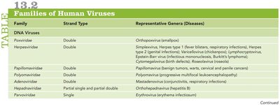
Viruses are classified based on their genetic material, structure, and host range. The following tables summarize the families of human viruses:
DNA Viruses
Family | Strand Type | Representative Genera (Diseases) |
|---|---|---|
Parvoviridae | Single | Erythrovirus (erythema infectiosum) |
Herpesviridae | Double | Simplexvirus, Varicellovirus, Cytomegalovirus, Epstein-Barr virus (herpes, chickenpox, mononucleosis, Burkitt's lymphoma) |
Papillomaviridae | Double | Papillomavirus (benign tumors, warts, cervical and penile cancers) |
Hepadnaviridae | Partial single and partial double | Orthohepadnavirus (hepatitis B) |
Adenoviridae | Double | Mastadenovirus (respiratory infections) |
RNA Viruses
Family | Strand Type | Representative Genera (Diseases) |
|---|---|---|
Picornaviridae | Single, + | Enterovirus (polio), Hepatovirus (hepatitis A), Rhinovirus (common cold) |
Flaviviridae | Single, + | Flavivirus (yellow fever), Hepacivirus (hepatitis C) |
Orthomyxoviridae | Single, - segmented | Influenzavirus (influenza) |
Retroviridae | Single, + | Lentivirus (HIV/AIDS) |
Reoviridae | Double | Rotavirus (diarrhea) |
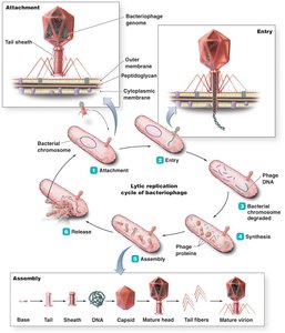
Viral Replication
Viruses depend on host organelles and enzymes to produce new virions. The lytic replication cycle typically results in the death and lysis of the host cell.
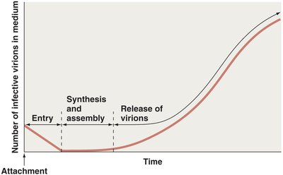
Stages of lytic replication: Attachment, Entry, Synthesis, Assembly, Release
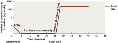
Pattern of Virion Abundance in Lytic Cycle
Virion numbers increase sharply after release by lysis.
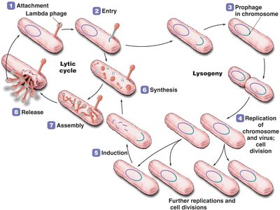
Lysogeny
Lysogeny is a modified replication cycle in which infected host cells grow and reproduce normally for generations before lysing.
Temperate phages can integrate their genome into the host chromosome as a prophage.
Lysogenic conversion occurs when phages carry genes that alter the phenotype of a bacterium.
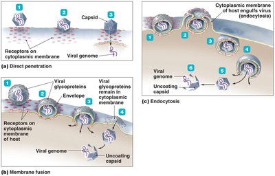
Replication of Animal Viruses
Animal viruses follow similar replication pathways as bacteriophages, with differences due to the presence of envelopes, eukaryotic cell structure, and lack of cell wall.
Attachment is mediated by glycoprotein spikes or other molecules.
Entry mechanisms include direct penetration, membrane fusion, and endocytosis.
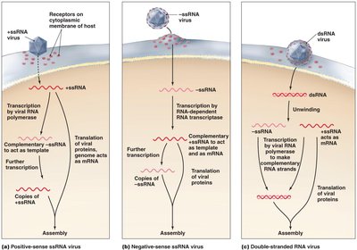
Synthesis of Animal Viruses
DNA viruses often replicate in the nucleus; RNA viruses in the cytoplasm.
Strategies depend on the type of nucleic acid.
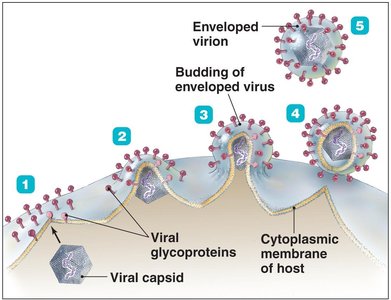
Assembly and Release of Animal Viruses
Most DNA viruses assemble in the nucleus; most RNA viruses in the cytoplasm.
Enveloped viruses cause persistent infections; naked viruses are released by exocytosis or lysis.
Pattern of Virion Abundance in Persistent Infections
Persistent infections show gradual increase in virion numbers.
Latency of Animal Viruses
Some animal viruses remain dormant in host cells for years, with no viral activity. Incorporation of provirus into host DNA is permanent.
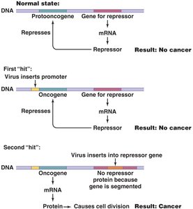
The Role of Viruses in Cancer
Viruses can contribute to cancer by carrying oncogenes, promoting host oncogenes, or interfering with tumor repression.
Neoplasia is uncontrolled cell division, forming tumors.
Viruses cause 20–25% of human cancers, including Burkitt’s lymphoma, Hodgkin’s disease, Kaposi’s sarcoma, and cervical cancer.
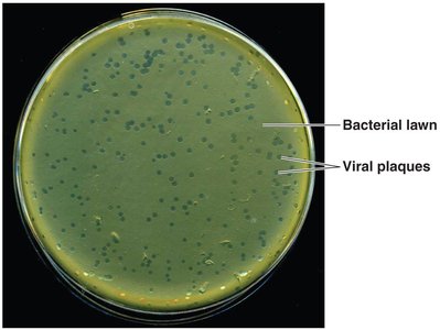
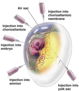
Culturing Viruses in the Laboratory
Viruses can be cultured in mature organisms, embryonated chicken eggs, or cell (tissue) cultures.
Viral plaques in bacterial lawns indicate viral infection.
Embryonated eggs provide a large, contaminant-free environment for viral growth.
Cell cultures can be diploid or continuous.
Other Parasitic Particles: Viroids and Prions
Viroids
Viroids are extremely small, circular pieces of RNA that are infectious and pathogenic in plants.
They lack a capsid and may appear linear due to hydrogen bonding.
Prions
Prions are proteinaceous infectious agents.
Cellular PrP has normal α-helices; prion PrP has disease-causing β-sheets.
Prion PrP induces conformational change in cellular PrP, leading to disease.
Prion diseases cause fatal neurological degeneration and spongiform encephalopathies (e.g., BSE, vCJD, kuru).
Prions are only destroyed by incineration or autoclaving in 1 N NaOH.
Are Viruses Alive?
There is debate about whether viruses are alive. Some consider them complex pathogenic chemicals, while others view them as the least complex living entities due to their ability to invade cells, control host machinery, and replicate.
Additional info: Academic context was added to clarify viral classification, replication, and the role of viruses in cancer, as well as to expand on viroids and prions.
