 Back
BackSynthesis, Packaging, and Release of Signaling Molecules: The Secretory Pathway in Eukaryotic Cells
Study Guide - Smart Notes
 Tailored notes based on your materials, expanded with key definitions, examples, and context.
Introduction to the Secretory Pathway
The secretory pathway is a highly conserved cellular process in eukaryotes, responsible for the synthesis, packaging, and release of signaling molecules such as hormones and neurotransmitters. This pathway involves a series of membrane-bound organelles and vesicular transport mechanisms that ensure proteins are correctly processed and delivered to their final destinations.
Professional Secretory Cells
Specialized Cell Types
Professional secretory cells, such as pancreatic exocrine cells and neurons, are used to study the mechanisms of protein sorting and packaging. These cells are specialized for the synthesis and regulated release of large quantities of signaling molecules.
Pancreatic exocrine cells: Secrete digestive enzymes.
Neurons: Release neurotransmitters for cell-to-cell communication.
Vesicular Transport and the Secretory Pathway
Overview of Vesicular Transport
Vesicular transport involves the movement of soluble proteins and membrane components between cellular compartments via transport vesicles. This process is essential for the proper functioning of the secretory pathway.
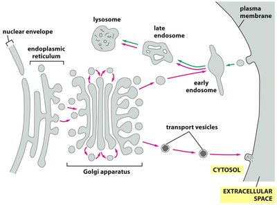
Endoplasmic Reticulum (ER): Site of protein synthesis and initial folding.
Golgi Apparatus: Modifies, sorts, and packages proteins for delivery.
Trans-Golgi Network (TGN): Final sorting station before proteins are sent to their destinations.
Secretory Vesicles: Store and release proteins via exocytosis.
Conservation Across Species
The secretory pathway is conserved from yeast to humans. Seminal studies in Saccharomyces cerevisiae (yeast) using temperature-sensitive mutants identified key proteins involved in vesicle formation, fusion, and cargo sorting.
sec1 (Munc18): Required for vesicle fusion with the plasma membrane.
sec7: Involved in TGN structure and vesicle formation.
sec61: Protein translocator complex for co-translational transport into the ER.
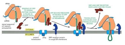
Protein Synthesis and Co-Translational Transport
Targeting Proteins to the ER
Proteins destined for secretion are synthesized by ribosomes bound to the rough ER. The signal recognition particle (SRP) directs the ribosome-mRNA complex to the ER membrane, where translation continues and the nascent polypeptide is translocated into the ER lumen.
Signal peptide: Short amino acid sequence that directs the protein to the ER.
SRP: Recognizes the signal peptide and pauses translation until docking at the ER.
Sec61 translocator: Channel through which the polypeptide enters the ER lumen.
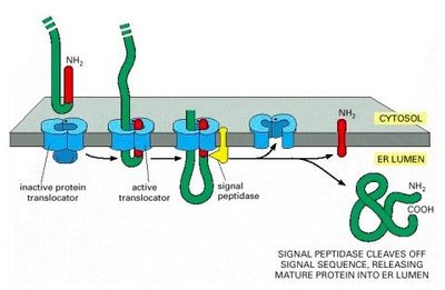
Signal Peptide Cleavage
Once the nascent protein enters the ER, the signal peptide is cleaved by signal peptidase, releasing the mature protein into the ER lumen.
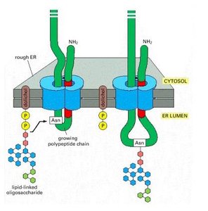
Protein Folding and Glycosylation in the ER
N-linked Glycosylation
During translation, a conserved oligosaccharide is added to asparagine residues of the nascent polypeptide (N-linked glycosylation). This modification is important for protein folding, stability, and quality control.
Calnexin and calreticulin: ER chaperones that bind incompletely folded glycoproteins, ensuring proper folding before exit from the ER.
Quality control: Proteins cannot leave the ER until folding is complete and all glucose residues are removed from the oligosaccharide.
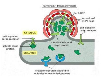
Vesicle Formation and Transport to the Golgi
ER Transport Vesicles
Properly folded proteins are packaged into COPII-coated vesicles that bud from the ER and are transported to the Golgi apparatus. Motor proteins move these vesicles along microtubules.
COPII coat: Protein complex that drives vesicle budding from the ER.
Vesicular tubular clusters: Intermediate structures formed during transport to the Golgi.
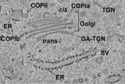
Golgi Apparatus: Processing and Sorting
Structure and Function
The Golgi apparatus consists of stacked cisternae with distinct cis (entry) and trans (exit) faces. Proteins undergo further modifications, such as glycosylation and proteolytic processing, and are sorted for delivery to their final destinations.
cis-Golgi: Receives vesicles from the ER.
trans-Golgi network (TGN): Sorting station for secretory vesicles, lysosomes, and the plasma membrane.
Clathrin and other coats: Mediate vesicle formation at the TGN for specific destinations.
Secretory Granule Formation and Exocytosis
Insulin Synthesis and Processing
Insulin is a classic example of a peptide hormone processed through the secretory pathway. In pancreatic β-cells, insulin is synthesized as preproinsulin, processed to proinsulin in the ER, and then cleaved to mature insulin in secretory granules.
Preproinsulin: Contains a signal peptide for ER targeting.
Proinsulin: Formed after signal peptide cleavage and folding in the ER.
Mature insulin: Produced after further cleavage in secretory vesicles, ready for regulated exocytosis.
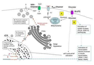
Exocytosis Mechanism
Secretory vesicles fuse with the plasma membrane in a Ca2+-dependent manner, releasing their contents into the extracellular space. This process is essential for hormone and neurotransmitter release.
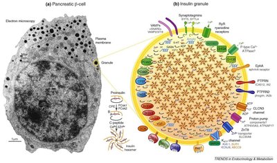
Dense core granules: Store peptide hormones like insulin.
Ca2+-dependent exocytosis: Triggered by an increase in intracellular calcium concentration.
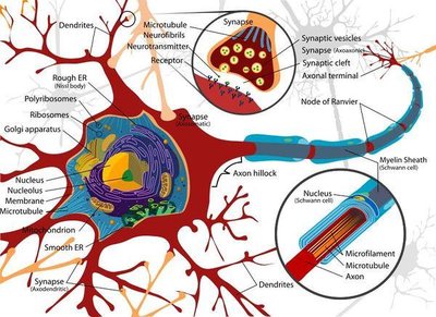
Neuronal Signaling and Neurotransmitter Release
Neuronal Structure and Function
Neurons are specialized cells for rapid communication via electrical and chemical signals. Neurotransmitters are synthesized, packaged into synaptic vesicles, and released at synapses to transmit signals to other cells.
Synaptic vesicles: Small vesicles that store neurotransmitters at the nerve terminal.
Exocytosis: Release of neurotransmitters into the synaptic cleft upon stimulation.
Comparison: Peptide vs. Amine Neurotransmitter Packaging
Feature | Peptide Hormones (e.g., Insulin) | Amine Neurotransmitters (e.g., Dopamine) |
|---|---|---|
Synthesis Site | Ribosomes (ER) | Cytosol (enzymatic) |
Processing | ER & Golgi (folding, glycosylation, cleavage) | Minimal processing |
Vesicle Type | Dense core granules | Small synaptic vesicles |
Packaging Mechanism | Co-translational import, vesicular transport | Vesicular transporter, ATPase-driven acidification |
Release | Ca2+-dependent exocytosis | Ca2+-dependent exocytosis |
Summary
The secretory pathway is fundamental to cell biology, enabling the synthesis, processing, and regulated release of signaling molecules. Understanding this pathway provides insight into essential physiological processes and the molecular basis of diseases such as diabetes and neurological disorders.
