 Back
BackThe Microbial World: Origins, Diversity, and Cell Structure
Study Guide - Smart Notes
 Tailored notes based on your materials, expanded with key definitions, examples, and context.
The Microbial World
Introduction to Microbiology
Microbiology is the study of microorganisms, which are life forms too small to be seen with the naked eye. Microbes include bacteria, archaea, some eukaryotes, and viruses. They are the most abundant and diverse forms of life on Earth, playing essential roles in ecosystems, biotechnology, and human health.
Microbe: Derived from 'micro' (small) and 'bios' (life), referring to microscopic organisms.
Biology: The scientific study of life, including the structure, function, growth, origin, evolution, and distribution of living organisms.
Importance: Microbes are crucial for nutrient cycling, environmental processes, and as model organisms in research.
Origins and Evolution of Life
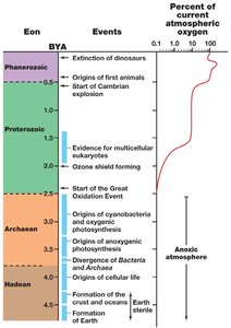
Formation of Earth and Early Life
The Earth formed approximately 4.6 billion years ago. Life is believed to have arisen around 3.6–3.8 billion years ago, as evidenced by ancient microfossils and stromatolites. Early life forms were likely chemolithotrophic, using inorganic molecules for energy, and later evolved photosynthetic capabilities.
Stromatolites: Layered sedimentary formations created by microbial mats, providing fossil evidence of early life.
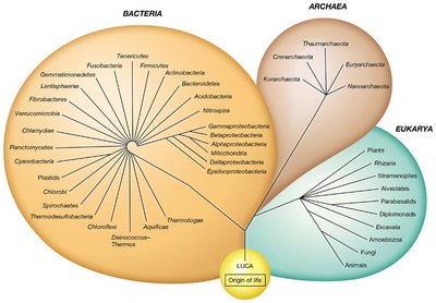
LUCA: The Last Universal Common Ancestor, from which all current life descends.
RNA World Hypothesis: Suggests that self-replicating RNA molecules were precursors to current life forms.
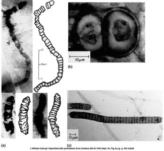
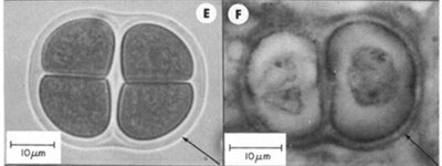
Fossil Evidence and Early Microbial Life
Fossilized remains of ancient microbes, such as cyanobacteria, have been found in rocks over 3.5 billion years old. These fossils provide insight into the evolution of cellular life and the development of oxygenic photosynthesis, which dramatically altered Earth's atmosphere.
Cyanobacteria: Photosynthetic bacteria responsible for the Great Oxidation Event (~2.4 billion years ago).
Stromatolites: Still found today, these structures are evidence of ancient microbial communities.
Classification and Diversity of Microorganisms
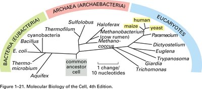
The Three Domains of Life
Modern classification divides all life into three domains based on ribosomal RNA (rRNA) sequence comparisons: Bacteria, Archaea, and Eukarya. This system reflects evolutionary relationships and fundamental differences in cell structure and genetics.
Bacteria: Prokaryotic, with peptidoglycan cell walls, ester-linked membrane lipids.
Archaea: Prokaryotic, lacking peptidoglycan, with ether-linked membrane lipids, often extremophiles.
Eukarya: Eukaryotic, with membrane-bound organelles and a true nucleus.
Taxonomy and Nomenclature
Taxonomy is the science of classifying organisms. It involves three main components: classification (grouping organisms), nomenclature (naming organisms), and identification (determining the identity of an organism). The binomial system, devised by Linnaeus, assigns each species a two-part Latin name (genus and species).
Example: Escherichia coli (abbreviated E. coli)
Phylogenetic classification: Based on evolutionary relationships, often using molecular data such as rRNA sequences.
Microbial Cell Structure and Function
Prokaryotic vs. Eukaryotic Cells
Microbial cells are classified as either prokaryotic or eukaryotic. Prokaryotes (Bacteria and Archaea) lack a true nucleus and membrane-bound organelles, while eukaryotes (Eukarya) possess these structures.
Prokaryotic cells: Simpler, usually smaller, with DNA in a nucleoid region.
Eukaryotic cells: More complex, with compartmentalized functions.
Prokaryotic Cell Morphology
Prokaryotes exhibit a variety of shapes and arrangements, including cocci (spherical), bacilli (rod-shaped), spirilla (spiral), and more. Some form multicellular structures or filaments.
Coccus: Spherical shape.
Bacillus: Rod-shaped.
Spirillum/Spirchete: Spiral-shaped, with spirochetes being more flexible.
Arrangements: Diplococci (pairs), streptococci (chains), staphylococci (clusters).
Cell Envelope Structure
The cell envelope includes the plasma membrane, cell wall, and, in some cases, an outer membrane or additional layers. The structure differs between Gram-positive and Gram-negative bacteria, which is the basis for the Gram stain.
Gram-positive: Thick peptidoglycan layer, teichoic acids, no outer membrane.
Gram-negative: Thin peptidoglycan, outer membrane with lipopolysaccharide (LPS), periplasmic space.
Archaeal cell walls: Lack peptidoglycan; may have pseudopeptidoglycan or S-layers.
Plasma Membrane and Transport
The plasma membrane is a selectively permeable barrier composed of a phospholipid bilayer with embedded proteins. It is essential for nutrient uptake, energy generation, and environmental sensing.
Transport mechanisms: Passive diffusion, facilitated diffusion, active transport, group translocation.
Membrane lipids: Bacteria and eukaryotes have ester-linked fatty acids; archaea have ether-linked isoprenoids.
Cytoskeleton and Internal Structures
Prokaryotes possess cytoskeletal proteins homologous to eukaryotic actin, tubulin, and intermediate filaments. These proteins are involved in cell division, shape maintenance, and chromosome segregation.
FtsZ: Tubulin homolog, forms a ring at the site of cell division.
MreB: Actin homolog, maintains rod shape.
Crescentin: Intermediate filament-like protein, involved in cell curvature.
Surface Structures
Many prokaryotes have external structures such as capsules, slime layers, S-layers, flagella, and pili. These structures aid in protection, attachment, motility, and genetic exchange.
Capsule: Well-organized, not easily removed, protects against phagocytosis.
Slime layer: Diffuse, unorganized, easily removed.
S-layer: Regular protein or glycoprotein layer, common in Archaea.
Flagella: Used for motility; rotation enables movement toward or away from stimuli (chemotaxis).
Pili (fimbriae): Shorter, used for attachment, movement, or conjugation (DNA transfer).
Microbial Nutrition and Growth
Essential Nutrients
Microorganisms require macronutrients (C, O, H, N, S, P, K, Ca, Mg, Fe) and micronutrients (Mn, Zn, Co, Mo, Ni, Cu) for growth. Carbon, energy, and electron sources define their nutritional types.
Autotrophs: Use CO2 as a carbon source.
Heterotrophs: Use organic molecules as carbon sources.
Phototrophs: Obtain energy from light.
Chemotrophs: Obtain energy from chemical compounds.
Lithotrophs: Use inorganic electron donors.
Organotrophs: Use organic electron donors.
Growth Factors
Some microbes require additional organic compounds called growth factors, such as amino acids, vitamins, purines, and pyrimidines, which they cannot synthesize themselves.
Uptake of Nutrients
Passive diffusion: Movement down a concentration gradient (e.g., O2, CO2).
Facilitated diffusion: Carrier-mediated, no energy required.
Active transport: Carrier-mediated, energy required (ATP or proton motive force).
Group translocation: Chemical modification during transport (e.g., PTS system for sugars).
Microbial Growth and Its Control
Growth Curve and Phases
Microbial growth in batch culture follows a characteristic curve with four phases: lag, exponential (log), stationary, and death.
Lag phase: Adaptation, no division.
Exponential phase: Rapid, constant division; balanced growth.
Stationary phase: Growth rate equals death rate; nutrient depletion or waste accumulation.
Death phase: Decline in viable cells.
Exponential growth equation:
Where is the number of cells at time , is the initial number of cells, and is the number of generations.
Environmental Factors Affecting Growth
Temperature: Psychrophiles, mesophiles, thermophiles, hyperthermophiles.
pH, oxygen, water activity, and other factors also influence microbial growth.
Control of Microbial Growth
Microbial populations can be controlled by physical and chemical methods:
Physical methods: Heat (autoclaving, pasteurization), filtration, radiation (UV, ionizing).
Chemical agents: Disinfectants (phenolics, alcohols, quaternary ammonium compounds, halogens), antiseptics.
Definitions: Sterilization (removal of all life), disinfection (removal of pathogens), sanitization (reduction to safe levels), antisepsis (prevention on living tissue).
Decimal reduction time (D value): Time required to kill 90% of microorganisms at a specific temperature.
Summary Table: Key Differences Among the Three Domains of Life
Feature | Bacteria | Archaea | Eukarya |
|---|---|---|---|
Membrane-bound nucleus | Absent | Absent | Present |
Cell wall | Peptidoglycan (murein) | No peptidoglycan; some have pseudopeptidoglycan | Varies; no peptidoglycan |
Membrane lipids | Ester-linked, straight-chain fatty acids | Ether-linked, branched isoprenoids | Ester-linked, straight-chain fatty acids |
Ribosomal RNA | 70S (16S rRNA) | 70S (16S rRNA) | 80S (18S rRNA) |
