 Back
BackThe Microbial World: Structure, Function, and Impact
Study Guide - Smart Notes
 Tailored notes based on your materials, expanded with key definitions, examples, and context.
The Microbial World
Introduction to Microorganisms
Microorganisms, or microbes, are life forms too small to be seen by the naked eye. They are highly diverse in form and function, inhabiting every environment that supports life. Most are single-celled, though some form complex structures or are multicellular. Microbes often live in communities, interacting with each other and their surroundings.
Definition: Microorganisms include bacteria, archaea, fungi, protozoa, algae, and viruses.
Importance: They are the oldest form of life and constitute a major fraction of Earth's biomass.
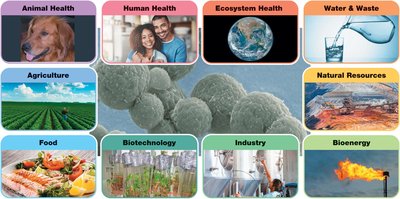
Applications: Microbes affect human life through infectious diseases, food and water safety, soil health, animal health, and fuel production.
Study Tools: Microscopy and cultivation techniques are essential for studying microbes.
Colony: A visible mass of microbial cells, usually containing millions or billions of cells.
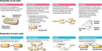
Structure and Activities of Microbial Cells
All cells share fundamental structural features, but there are key differences between prokaryotic and eukaryotic cells. The cell is a living compartment that interacts with its environment and other cells.
Cytoplasmic (cell) membrane: Barrier separating the cytoplasm from the external environment.
Cytoplasm: Aqueous mixture of macromolecules, small organics, ions, and ribosomes.
Ribosomes: Structures responsible for protein synthesis.
Cell wall: Present in some microbes, confers structural strength.
Prokaryotes (Bacteria and Archaea) lack membrane-bound organelles and a nucleus, while eukaryotes (plants, animals, algae, protozoa, fungi) contain organelles and have DNA enclosed in a nucleus.
Genome: The full set of genes in a cell.
Eukaryotic DNA: Linear chromosomes within a nucleus; larger and more complex.
Prokaryotic DNA: Typically a single circular chromosome in the nucleoid region; may have plasmids.
Microbial cells perform various activities:
Metabolism: Chemical transformation of nutrients, catalyzed by enzymes.
Transcription: DNA information converted to RNA.
Translation: RNA used by ribosomes to synthesize proteins.
DNA replication: Copying the genome.
Motility: Movement through self-propulsion.
Differentiation: Formation of specialized cells.
Intercellular communication: Response to chemical signals from other microbes.
Evolution: Genetic changes transferred to offspring over time.
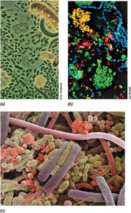
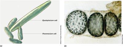
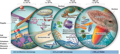
Cell Size and Morphology
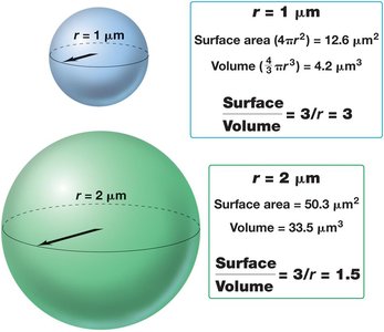
Cell morphology refers to the size and shape of cells. Prokaryotes range from 0.2 µm to over 600 µm in diameter, while eukaryotic cells are typically 5–100 µm in length. Cell size affects surface-to-volume ratios, which influence nutrient exchange and growth rates.
Surface-to-volume ratio: Smaller cells have a higher ratio, supporting greater nutrient and waste exchange per unit volume.
Major morphologies: Cocci (spherical), rods (cylindrical), spirilla (spiral), spirochetes (rigid spiral), appendaged, irregular/asymmetrical, filamentous.
An Introduction to Microbial Life
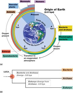
Microbial life is classified into three domains: Bacteria, Archaea, and Eukarya. Each domain has unique characteristics and evolutionary histories.
Bacteria: Prokaryotes, usually undifferentiated single cells, with 80+ phylogenetic lineages.
Archaea: Prokaryotes, often associated with extreme environments, with 12+ phyla.
Eukarya: Includes plants, animals, fungi; first were unicellular, at least six kingdoms.
Viruses: Obligate parasites, not cells, replicate only within host cells, classified by structure, genome, and host specificity.
Microorganisms and the Biosphere
Microbes have played a crucial role in Earth's history and the biosphere. The first cells appeared between 3.8 and 4.3 billion years ago, and the atmosphere was anoxic until about 2.6 billion years ago. Extremophiles live in habitats too harsh for other life forms, such as hot springs, glaciers, high salt, acidity, alkalinity, and pressure.
LUCA: Last universal common ancestor, from which Bacteria, Archaea, and Eukarya descended.
Microbial ecology: Study of how microbes affect animals, plants, and global ecosystems.
Global biomass: Microbial cells contribute significantly to Earth's carbon, nitrogen, and phosphorus cycles.

The Impact of Microorganisms on Human Society
Microorganisms can be both beneficial and harmful to humans. They are agents of disease, play roles in food and agriculture, and are valuable for human products, energy generation, and environmental clean-up.
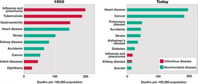
Disease agents: Control of infectious diseases, vaccination, antibiotic therapy, water and wastewater treatment, food safety.
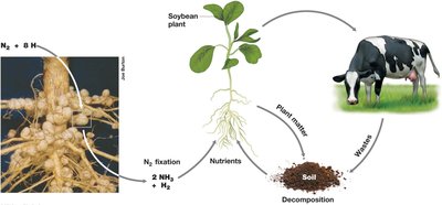
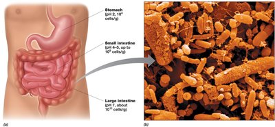
Agriculture: Nitrogen-fixing bacteria, cellulose-degrading microbes, gut microbiome, synthesis of vitamins and nutrients.
Food: Can cause spoilage and foodborne disease, but also improve food safety and preservation through fermentation.
Industry: Use of microbes in pharmaceuticals, brewing, biotechnology, biofuel production, wastewater treatment, bioremediation, and biofilms.
Microscopy and the Origins of Microbiology
Light Microscopy and the Discovery of Microorganisms
Microbiology began with the invention of the microscope. Robert Hooke first described microbes, and Antoni van Leeuwenhoek was the first to see bacteria. Light microscopes use visible light to illuminate samples, with magnification and resolution as key properties.
Types: Bright-field, phase-contrast, differential interference contrast, dark-field, fluorescence.
Compound light microscope: Uses objective and ocular lenses; total magnification is the product of both.
Bright-field scope: Visualizes specimens based on contrast differences.
Improving Contrast in Light Microscopy
Staining increases contrast for bright-field microscopy. Basic dyes bind strongly to negatively charged cell components. Differential stains, such as the Gram stain, render different kinds of cells different colors based on cell wall structure.
Simple stain: Uses dried cells and basic dyes.
Gram stain: Gram-positive bacteria appear purple-violet; Gram-negative bacteria appear pink.
Phase-contrast microscopy: Amplifies differences in refractive index; dark cells on a light background.
Dark-field microscopy: Light scattered by specimen; image appears light on a dark background.
Fluorescence microscopy: Visualizes specimens that fluoresce; cells appear to glow on a black background.
Imaging Cells in Three Dimensions
Differential interference contrast (DIC) microscopy uses polarized light to give structures a three-dimensional appearance. Confocal scanning laser microscopy (CSLM) generates three-dimensional images by focusing a laser on single layers of the specimen.
Probing Cell Structure: Electron Microscopy
Electron microscopes use electrons instead of visible light to image cells and structures. Transmission electron microscopes (TEM) have greater resolving power and enable visualization at the molecular level. Scanning electron microscopes (SEM) visualize surfaces of specimens.
Microbial Cultivation Expands the Horizon of Microbiology
Pasteur and Spontaneous Generation
Louis Pasteur disproved the theory of spontaneous generation using the swan-necked flask experiment. He discovered that fermentation was a biological process and developed vaccines for anthrax, fowl cholera, and rabies.
Koch, Infectious Disease, and Pure Cultures
Robert Koch demonstrated the link between microbes and infectious diseases, identified causative agents, and developed Koch's postulates to link cause and effect. He developed solid media for obtaining pure cultures.
Discovery of Microbial Diversity
Sergei Winogradsky demonstrated that specific bacteria are linked to biogeochemical transformations and proposed chemolithotrophy. Martinus Beijerinck developed enrichment culture techniques and isolated the first aerobic nitrogen-fixing bacterium.
Molecular Biology and the Unity and Diversity of Life
Molecular Basis of Life
Bacteria are excellent models for studying the fundamental nature of life. Key discoveries include genetic transfer in bacteria, DNA as the genetic material, and the structure of DNA.
Woese and the Tree of Life
Carl Woese used ribosomal RNA sequences to infer evolutionary relationships, discovering the domain Archaea. The phylogenetic tree depicts the evolutionary history of all cells, with three domains and LUCA as the root.
Metagenomics: Allows recovery of microbial genomes from environmental DNA samples.
Additional info: These notes cover foundational concepts in microbiology, including microbial diversity, cell structure and function, microscopy, microbial cultivation, and molecular biology. They are suitable for exam preparation and provide a comprehensive overview of the microbial world.
