 Back
BackThe Molecular Basis of Genetic Material: DNA, RNA, and Protein Structure
Study Guide - Smart Notes
 Tailored notes based on your materials, expanded with key definitions, examples, and context.
Bacterial Genome and the Discovery of DNA as Genetic Material
Introduction to the Genome
The genome is the complete set of genetic material present in a cell or virus.
In bacteria and archaea, the genome is typically haploid (one set of genes), while eukaryotes are usually diploid (two sets).
The genotype refers to the specific set of genes an organism possesses, and the phenotype is the collection of observable characteristics resulting from gene expression.
Mutation – heritable change in genetic material
Historical Experiments Demonstrating DNA as Genetic Material
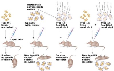
Griffith’s Transformation Experiments (1928)
Frederick Griffith discovered the phenomenon of transformation using Streptococcus pneumoniae. He observed that nonvirulent bacteria could be transformed into virulent forms, suggesting the presence of a 'transforming principle.' Initially, he hypothesized this molecule was a protein.
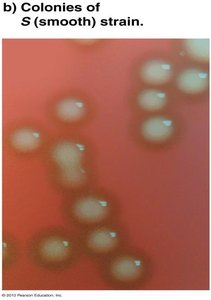
S (smooth) strain: Virulent, produces a polysaccharide capsule, resists host immunity, and causes death in mice.
The III-S strain synthesized a polysaccharide capsule that protected itself from the host’s immunity, resulting in the death of the host
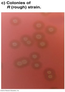
R (rough) strain: Nonvirulent, lacks capsule, and is eliminated by the host immune system.
I-R strain has a mutation that leads to no protective capsule and was defeated by the host's immune system.
Mixing heat-killed S strain with live R strain resulted in the transformation of R to S, causing mouse death.
Concluded that IIR bacteria has somehow been transformed into smooth, virulent IIIS bacteria by interacting with IIIS strain. Griffith was wrong to propose this was due to proteins (no experimental evidence to support this).
The IIIS strain bacteria had been killed, the DNA had survived the heating process and was taken up by the II-R strain bacteria.
Avery, MacLeod, and McCarty’s Experiments (1944)
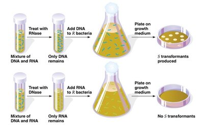
These scientists identified DNA as the 'transforming principle' by isolating nucleic acids from S strain bacteria and demonstrating that only DNA (not RNA or protein) could transform R strain bacteria into S strain.
Enzymatic removal of DNA prevented transformation, confirming DNA's role.
Also worked with Streptococcus pneumoniae type IIIS Separated nucleic acids (DNA, RNA) from remaining cell components
Concluded that “IIIS” DNA was the transforming agent responsible for Griffith’s results (not RNA).
Hershey-Chase Experiment (1953)
Alfred Hershey and Martha Chase used bacteriophages (viruses that infect bacteria) to confirm that DNA, not protein, is the genetic material. They labeled phage DNA with radioactive phosphorus (32P) and protein with radioactive sulfur (35S). Only DNA entered the bacterial cells and directed the production of new phages.
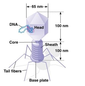
Bacteriophage structure: Composed of DNA and protein.
Virus that attacks bacteria and replicates by invading a living cell and using the host cell’s molecular machinery. Some function as parasitoids
Components: DNA & protein
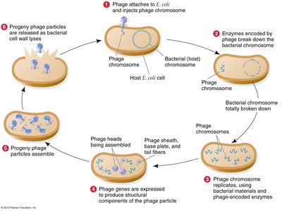
Lytic cycle: Viral DNA replicates independently of host DNA, leading to cell lysis.
Viral DNA exists as a separate free floating molecule within the bacterial cell and replicates separately from the host bacterial DNA. - E. coli T4 bacteriophage replicates this way
Lysogenic cycle: Viral DNA integrates into the host genome and can remain dormant.
the viral DNA is integrated into the host genome. The viral genome is dormant and can be passed to the next bacterial generation. UV or some chemical triggers can activate the genome and causing proliferation of new phages .
In both cases the virus/phage replicates using the host
DNA machinery.
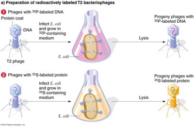
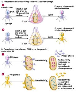
T2 bacteriophage is composed of DNA and proteins:
Set-up two treatments:
In one, labeled DNA with isotope 32P
In another, labeled protein with isotope 35S
Infected E. coli bacteria with two types of labeled T2
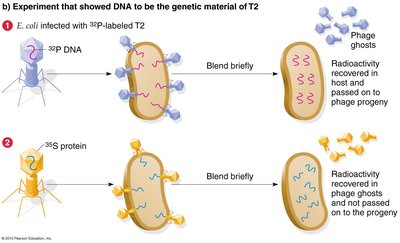
32P is discovered within the bacteria and T2 phage progeny, whereas 35S is not found within the bacteria but released with phage ghosts.
Conclusion:
Griffith 1928: One genetic strain can transform another to have a new trait. (He incorrectly concluded the transforming agent is a protein)
Avery 1944: DNA (not RNA) is transforming agent.
Hershey-Chase 1953: DNA (not protein) is the genetic material.
Chemical Structure of DNA and RNA
Nucleic Acids: DNA and RNA
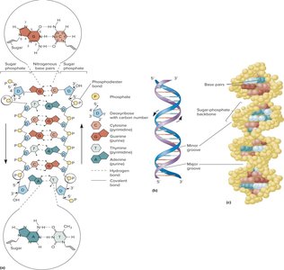
Both DNA and RNA are polymers of nucleotides linked by phosphodiester bonds. They differ in their nitrogenous bases, sugars, and strand structure.
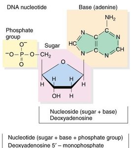
DNA: Contains deoxyribose sugar, bases adenine (A), guanine (G), cytosine (C), and thymine (T); usually double-stranded.
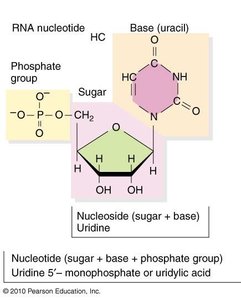
RNA: Contains ribose sugar, bases adenine (A), guanine (G), cytosine (C), and uracil (U); usually single-stranded.
Polynucleotide Chains and Phosphodiester Bonds
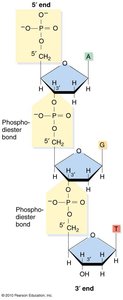
Nucleotides are joined by strong covalent phosphodiester bonds between the 5' phosphate of one nucleotide and the 3' hydroxyl of the next. This gives DNA and RNA strands polarity (5' to 3' directionality) and remarkable stability.
Nucleotides are linked by phosphodiester bonds to form polynucleotides.
Phosphodiester bond: Covalent bond between the phosphate group (attached to 5’ carbon) of one nucleotide and the 3’ carbon of the sugar of another nucleotide. This bond is very strong, and for this reason DNA is remarkably stable:
DNA can be boiled or autoclaved without degrading
Forensic science is an obvious application of its durability
Why would it be important for DNA to be stable?
5’ and 3’: The ends of the DNA or RNA chain are not the same:
One end of the chain has a 5’ carbon (with a phosphate group on it) and the other end has a 3‘ carbon (hydroxyl group on it).
Therefore, a DNA molecule has polarity.
a DNA strand is a polynucleotides chain
3 Parts of a Nuc:
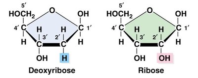
Pentose (5-carbon) sugar:
DNA = deoxyribose
RNA = ribose (compare 2’ carbons
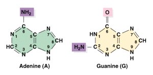
Nitrogenous base:
attached to 1’ carbon
Include heterocyclic ring
Purines: Adenine, Guanine
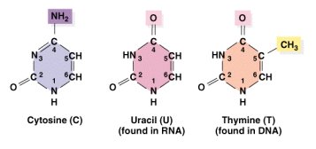
Pyrimidines: Cytosine, Thymine (DNA) Uracil (RNA)
Phosphate group attached to 5‘carbon of the sugar
and 3’ of another
Base Pairing and Double Helix Structure
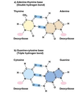
Bases from opposite DNA strands pair via hydrogen bonds: A with T (2 bonds), G with C (3 bonds). This pairing is essential for the double helix structure, which consists of two antiparallel strands wound in a right-handed helix. The sugar-phosphate backbone is on the outside, and the bases are oriented inward.
Chargaff’s rules: Amount of A = T, G = C; %GC or %AT content varies among organisms.
Major and minor grooves: Result from the geometry of the double helix.
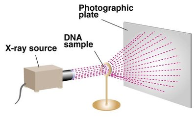
Experimental Evidence for DNA Structure
Watson and Crick (1953) proposed the double helix model based on Chargaff’s base composition studies and X-ray diffraction data from Rosalind Franklin and Maurice Wilkins.
6 Main Features of the DNA Double Helix
Two polynucleotide chains wound in a right-handed (clockwise) double-helix.
Nucleotide chains are anti-parallel: 5’ → 3’ 3’ → 5‘
Sugar-phosphate backbones are on the outside of the double helix, and the bases are oriented towards the central axis.
Complementary base pairs from opposite strands are bound together by weak hydrogen bonds.
A pairs with T (2 H-bonds), and G pairs with C (3 H-bonds).
e.g., 5’-TATTCCGA-3’ 3’-ATAAGGCT-5’
Base pairs are 0.34 nm apart. One complete turn of the helix requires 3.4 nm (10 bases/turn).
Sugar-phosphate backbones are not equally-spaced, resulting in major and minor grooves.
RNA Structure
RNA is typically single-stranded but can form secondary structures such as hairpin loops through complementary base pairing. It contains ribose sugar and uracil instead of thymine.
Polymer of nucleotides.
• Contains the bases adenine, guanine, cytosine, and uracil.
• Sugar is ribose.
• The nucleotides are joined by phosphodiester bonds, just as they do in
DNA.
Most RNA molecules single stranded; some double stranded.
When single stranded it can coil back on itself to form secondary
structures such as hairpin loops, with complementary base
paring
Protein Structure and Amino Acids
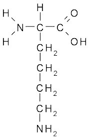
Amino Acids
Amino acids are the building blocks of proteins. Each has a central (alpha) carbon bonded to an amino group (N), a carboxyl group (C) , a hydrogen atom, and a variable side chain (R group).
Amino acids can be polar, nonpolar, or charged depending on their side chains.
Peptide Bonds and Protein Structure
Proteins are polymers of amino acids linked by peptide bonds, which form between the carboxyl group of one amino acid and the amino group of the next, releasing water (condensation reaction). The sequence of amino acids determines the protein's structure and function.

Summary Table: Key Differences Between DNA and RNA
Feature | DNA | RNA |
|---|---|---|
Sugar | Deoxyribose | Ribose |
Bases | A, T, G, C | A, U, G, C |
Strandedness | Double-stranded | Single-stranded (usually) |
Stability | Very stable | Less stable |
Main Function | Genetic information storage | Information transfer, catalysis |
Additional info: The stability of DNA is crucial for the preservation of genetic information across generations, while the relative instability of RNA allows for its roles in transient information transfer and regulation.
