 Back
BackTissue Level of Organization: Epithelial and Connective Tissues
Study Guide - Smart Notes
 Tailored notes based on your materials, expanded with key definitions, examples, and context.
Tissue Level of Organization
Introduction to Tissues
Tissues are groups of cells that work together to perform specialized functions in the body. The study of tissues is known as histology. There are four primary types of tissues: epithelial, connective, muscle, and nervous tissue, each with distinct roles and characteristics.
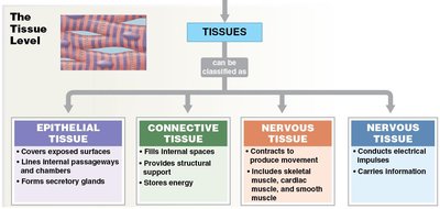
Epithelial tissue: Covers surfaces, lines cavities, forms glands.
Connective tissue: Provides structural support, fills spaces, stores energy.
Muscle tissue: Contracts to produce movement.
Nervous tissue: Conducts electrical impulses, carries information.
Epithelial Tissue
Structure and Functions
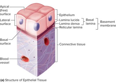
Epithelial tissue consists of closely packed cells with minimal extracellular matrix. It forms protective barriers, controls permeability, and is involved in absorption, secretion, and sensory reception. Epithelial cells exhibit polarity, with distinct apical, basal, and lateral surfaces.
Physical protection: Shields underlying tissues from mechanical and chemical injury.
Control permeability: Regulates entry and exit of substances.
Absorption and secretion: Specialized for uptake and release of molecules.
Sensory reception: Contains nerve endings for sensation.
Regeneration: Rapidly replaces damaged cells.
Basement Membrane
The basement membrane is a specialized extracellular structure that anchors epithelial cells to underlying connective tissue. It consists of two layers: the basal lamina and the reticular lamina, composed of collagen, glycoproteins, and proteoglycans.

Basal lamina: Secreted by epithelial cells, provides support and filtration.
Reticular lamina: Produced by connective tissue, adds strength.
Cell Shapes and Arrangements
Epithelial cells are classified by their shape and the number of layers:
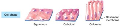
Squamous: Flat, thin cells.
Cuboidal: Cube-shaped cells.
Columnar: Tall, column-like cells.
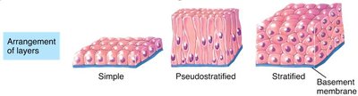
Simple: Single layer.
Stratified: Multiple layers.
Pseudostratified: Appears layered but all cells touch the basement membrane.
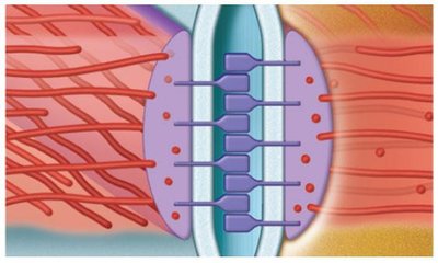
Intercellular Connections (Junctions)
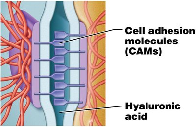
Intercellular junctions are specialized structures that connect epithelial cells, providing mechanical strength and regulating permeability.
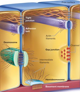
Desmosomes: Join cytoskeletons of adjacent cells, resist mechanical stress.
Hemidesmosomes: Anchor cells to the basement membrane.
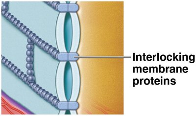
Tight junctions: Seal cells together, prevent leakage of fluids.
Adhesion belts: Continuous bands that resist separation.
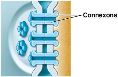
Gap junctions: Channels for communication and passage of ions/molecules.
Types of Epithelia
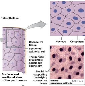
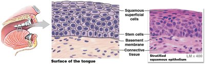
Squamous Epithelium
Squamous epithelium consists of flat cells and can be simple or stratified. Simple squamous is specialized for diffusion and filtration, while stratified squamous provides protection against abrasion.
Simple squamous: Found in peritoneum, capillaries, alveoli, kidneys.
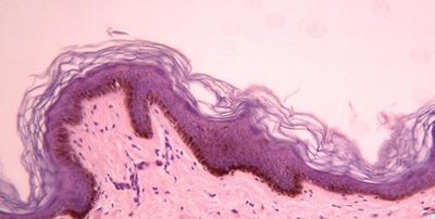
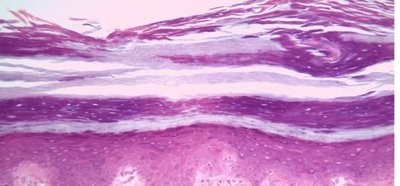
Stratified squamous: Forms skin, lines mouth, esophagus, vagina.
Keratinized: Epidermis, resists dehydration.
Nonkeratinized: Moist surfaces, e.g., oral cavity.
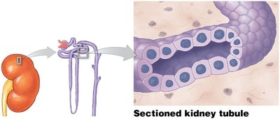
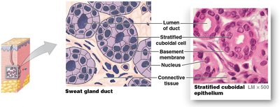
Cuboidal Epithelium
Cuboidal epithelium consists of cube-shaped cells. Simple cuboidal is involved in secretion and absorption, while stratified cuboidal is rare and provides protection.
Simple cuboidal: Lines glands, ducts, kidney tubules.
Stratified cuboidal: Found in sweat glands, mammary glands.
Transitional Epithelium
Transitional epithelium is specialized for stretching and recoiling, found in the urinary bladder and ureters.
Function: Protection and distention.
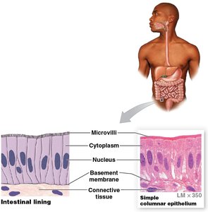
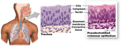
Columnar Epithelium
Columnar epithelium consists of tall, column-like cells. Simple columnar is specialized for absorption and secretion, often with microvilli or cilia. Pseudostratified columnar appears layered but all cells touch the basement membrane. Stratified columnar is rare and provides protection and secretion.
Simple columnar: Lines digestive, respiratory, reproductive tracts.
Pseudostratified columnar: Found in upper respiratory tract, ducts.
Stratified columnar: Lines urethra, salivary glands.

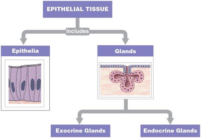
Glandular Epithelium
Glandular epithelia are specialized for secretion. Glands can be classified as endocrine (release hormones into interstitial fluid) or exocrine (release secretions into ducts).
Unicellular glands: Single cells, e.g., goblet cells.
Multicellular glands: Simple (single duct) or compound (branched ducts).

Modes of Exocrine Secretion
Mode | Description | Example |
|---|---|---|
Merocrine | Product released by exocytosis | Salivary glands |
Apocrine | Apical cytoplasm released with product | Mammary glands |
Holocrine | Entire cell bursts, releasing product | Sebaceous glands |
Connective Tissue
General Structure and Functions
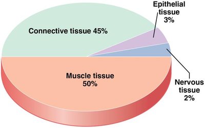
Connective tissue is the most abundant tissue type, characterized by few cells and abundant extracellular matrix. It binds, supports, protects, and insulates organs, connects tissues, stores energy, and defends against microorganisms.
Specialized cells: -blast (immature), -cyte (mature), -clast (matrix breakdown).
Extracellular protein fibers: Collagen, elastin, reticular.
Ground substance: Fluid, semifluid, gelatinous, fibrous, calcified.
Matrix: Ground substance + fibers.
Connective tissue contains various cell types, including fibroblasts, adipocytes, mast cells, erythrocytes, leukocytes, macrophages, and platelets, each with specialized functions.
Comparison: Epithelial vs. Connective Tissue
Feature | Epithelial Tissue | Connective Tissue |
|---|---|---|
Cellularity | Many cells, tightly packed | Few cells, widely spaced |
Extracellular Matrix | Minimal | Abundant |
Blood Supply | Avascular | Rich blood supply |
Epithelial cells are always bound to underlying connective tissue via the basement membrane.
Additional info: These notes provide foundational knowledge for histology and tissue organization, essential for understanding cell structure and function in advanced biology and health sciences.
