 Back
BackTranslation: From RNA to Protein – The Genetic Code and Protein Synthesis
Study Guide - Smart Notes
 Tailored notes based on your materials, expanded with key definitions, examples, and context.
Translation: RNA to Protein
Overview of Translation
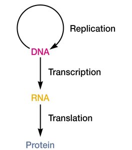
Translation is the process by which the genetic code carried by messenger RNA (mRNA) is decoded to produce a specific sequence of amino acids, resulting in the synthesis of a polypeptide (protein). This process is a fundamental aspect of the central dogma of molecular biology, which describes the flow of genetic information from DNA to RNA to protein.
Translation is the conversion of the nucleotide sequence of mRNA into the amino acid sequence of a protein.
It is carried out by the ribosome, which reads the mRNA from the 5' to 3' direction and synthesizes the polypeptide from the N-terminus to the C-terminus.
Translation is directed by the sequence of codons in the mRNA.
The Genetic Code
Structure and Properties of the Genetic Code
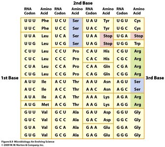
The genetic code is a set of rules by which information encoded in mRNA sequences is translated into proteins by living cells. Each group of three nucleotides (codon) in mRNA corresponds to a specific amino acid or a stop signal during protein synthesis.
Triplet Code: Each codon consists of three nucleotides, allowing for 64 possible codons (43 = 64).
Start Codon: AUG (codes for methionine) signals the start of translation.
Stop Codons: UAA, UAG, and UGA signal the termination of translation.
Degeneracy: Most amino acids are encoded by more than one codon (61 sense codons for 20 amino acids).
Universality: The genetic code is nearly universal among all organisms, with minor exceptions (e.g., mitochondrial genes).
Non-overlapping: Codons are read one after another without overlap.
tRNA and Decoding the Genetic Code
tRNA Structure and Function
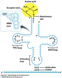
Transfer RNA (tRNA) molecules serve as adaptors that match specific codons in the mRNA with their corresponding amino acids during translation. Each tRNA has an anticodon region that base-pairs with the codon on the mRNA and an acceptor stem where the corresponding amino acid is attached.
Anticodon: A sequence of three bases on tRNA that is complementary to the mRNA codon.
Isoacceptors: Different tRNAs that accept the same amino acid but have different anticodons.
Wobble Hypothesis: The third base of the codon (and the first base of the anticodon) can form non-standard base pairs, allowing some tRNAs to recognize multiple codons.
Wobble Base Pairing
The wobble hypothesis explains how a single tRNA can recognize more than one codon. The first position of the anticodon (5' end) can pair with more than one base at the third position of the codon (3' end), increasing the efficiency of translation.
Watson-Crick Base Pairing: Standard base pairing (A-U, G-C) occurs at the first two positions of the codon.
Wobble Position: Non-standard pairing occurs at the third codon position, allowing flexibility.
Aminoacyl-tRNA Synthetases
Charging tRNAs with Amino Acids
Aminoacyl-tRNA synthetases are enzymes that attach the correct amino acid to its corresponding tRNA. Each amino acid has its own synthetase, which recognizes all compatible tRNAs (isoacceptors) for that amino acid.
Specificity: Ensures that the genetic code is translated accurately.
Reaction: Amino acid + tRNA + ATP → aminoacyl-tRNA + AMP + PPi
The Ribosome and Sites of Translation
Structure of the Prokaryotic Ribosome
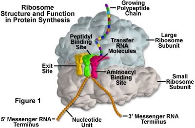
The ribosome is a complex molecular machine composed of ribosomal RNA (rRNA) and proteins. In prokaryotes, the ribosome consists of a small (30S) and a large (50S) subunit, which together form the functional 70S ribosome.
30S Subunit: Responsible for decoding the mRNA and aligning the tRNA anticodon.
50S Subunit: Catalyzes peptide bond formation.
Sites: A (aminoacyl), P (peptidyl), and E (exit) sites coordinate tRNA binding and movement.
Stages of Translation
Initiation
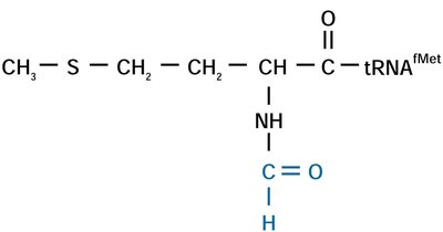
Translation initiation involves the assembly of the ribosome on the mRNA at the correct start codon. In bacteria, the Shine-Dalgarno sequence on the mRNA base-pairs with the 16S rRNA of the 30S subunit to position the ribosome correctly. The initiator tRNA carries N-formylmethionine (fMet) in bacteria.
Initiation Factors: IF1, IF2, and IF3 assist in ribosome assembly and initiator tRNA placement.
Start Codon: Typically AUG, recognized by initiator tRNA.
Elongation
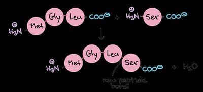
During elongation, amino acids are added one by one to the growing polypeptide chain. The ribosome moves along the mRNA, and tRNAs bring the appropriate amino acids to the A site. Peptide bonds are formed at the P site, and the empty tRNA exits from the E site.
Elongation Factors: EF-Tu, EF-G, and EF-Ts facilitate tRNA entry, translocation, and recycling.
Directionality: mRNA is read 5' to 3'; polypeptide is synthesized N-terminus to C-terminus.
Termination
Translation terminates when a stop codon (UAA, UAG, or UGA) is encountered. Release factors (RF1, RF2, RF3) recognize stop codons and promote the release of the completed polypeptide from the ribosome.
Release Factors: Bind to the ribosome and catalyze the release of the polypeptide chain.
Antibiotics Targeting Translation
Mechanisms of Inhibition
Several antibiotics inhibit bacterial translation by targeting different steps or components of the process. These drugs are important tools in medicine and research.
Inhibitor | Action |
|---|---|
Streptomycin | Causes mRNA misreading & inhibits chain initiation |
Puromycin | Aminoacyl-tRNA analog that causes premature chain termination |
Tetracycline | Inhibits binding of aminoacyl-tRNA to 30S subunit |
Fusidic acid | Blocks elongation by preventing dissociation of EF-G-GDP from ribosome |
Chloramphenicol | Inhibits peptidyl transferase activity in 50S subunit |
Erythromycin | Inhibits translocation by 50S subunit |
Summary Table: The Genetic Code
The following table summarizes the codons and their corresponding amino acids, including start and stop codons.
1st Base | 2nd Base | 3rd Base | Amino Acid |
|---|---|---|---|
U | U | U | Phe |
U | U | C | Phe |
U | U | A | Leu |
U | U | G | Leu |
U | A | A | Stop |
U | A | G | Stop |
A | U | G | Met (Start) |
G | G | G | Gly |
Additional info: The above notes integrate foundational concepts from the central dogma, the structure and function of the genetic code, tRNA, ribosomes, and the mechanism of translation, as well as the action of antibiotics that target translation. This content is essential for understanding gene expression and protein synthesis in microbiology.
