 Back
BackViral Structure, Classification, Replication, Pathogenesis, and Antiviral Strategies
Study Guide - Smart Notes
 Tailored notes based on your materials, expanded with key definitions, examples, and context.
Viral Structure and the Virosphere
Introduction to Viruses
Viruses are the most abundant biological entities on Earth, existing in diverse environments from deep caves to oceans. They are obligate intracellular parasites, meaning they require a host cell to replicate. Viruses are not considered living organisms because they lack cellular machinery and metabolic processes.
Definition: An infectious, obligate intracellular parasite comprising genetic material (DNA or RNA), often surrounded by a protein coat, and sometimes a lipid membrane.
Virosphere: The collective term for all viruses.
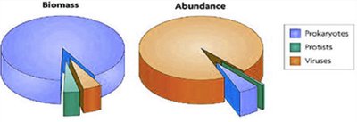
Abundance: There are approximately 1030 bacterial viruses (bacteriophages) alone, with a total viral biomass exceeding that of all elephants on Earth.
Visibility: Viruses are extremely small and can only be visualized with an electron microscope.
Structural Components of Viruses
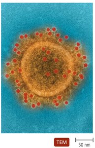
Viruses are composed of genetic material, a protein capsid, and in some cases, a lipid envelope. The structure determines their classification and host specificity.
Genome: DNA or RNA, which may be single-stranded, double-stranded, linear, segmented, or circular.
Capsid: Protein shell composed of repeating units called capsomeres.
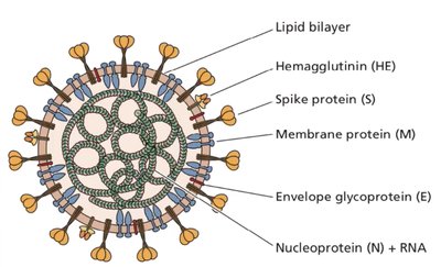
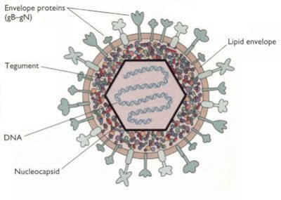
Envelope: Some viruses possess a lipid membrane derived from the host cell, embedded with glycoproteins (spikes).
Virion: The complete, infectious virus particle outside a host cell.
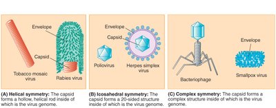
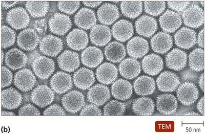
Virus Symmetry and Morphology
Viruses exhibit different structural symmetries, which are important for classification and function.
Helical Symmetry: Capsid forms a hollow, helical rod (e.g., Tobacco mosaic virus).
Icosahedral Symmetry: Capsid forms a 20-sided structure (e.g., Poliovirus, Herpes simplex virus).
Complex Symmetry: More elaborate structures, such as bacteriophages.
Classification and Taxonomy of Viruses
Taxonomic Organization
Viruses are classified based on their genetic material, envelope presence, and capsid structure. The International Committee on Taxonomy of Viruses (ICTV) provides a standardized nomenclature.
Family: Ends with -viridae (e.g., Flaviviridae).
Genus: Ends with -virus (e.g., Flavivirus).
Species: Not italicized; common names are used (e.g., yellow fever virus).
Classification Criteria: Genome type, envelope presence, capsid structure, host range, and tissue tropism.
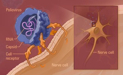
Host Range and Specificity
Viruses typically have a narrow host range, determined by the interaction between viral surface proteins and host cell receptors. Tissue tropism refers to the specific cell types within a host that a virus can infect.
Example: Poliovirus infects only humans, while rabies virus can infect most warm-blooded animals.
Mechanism: Viral attachment proteins (spikes) bind to specific receptors on host cells, mediating entry.
Viral Replication Cycles
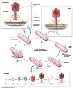
Bacteriophage Replication
Bacteriophages (viruses that infect bacteria) can undergo lytic or lysogenic replication cycles.
Lytic Cycle: Involves attachment, entry, degradation of host DNA, synthesis of viral components, assembly, and release of new virions, often resulting in host cell lysis.
Lysogenic Cycle: Viral genome integrates into the host chromosome (prophage), replicates with the host, and can later be induced to enter the lytic cycle.
Animal Virus Replication
Animal viruses attach to host cells via spikes on the capsid or envelope. Entry mechanisms include membrane fusion (enveloped viruses) or endocytosis (naked viruses). Replication involves uncoating, genome replication, protein synthesis, assembly, and release.
Enveloped Viruses: Often exit the cell by budding, preserving host cell integrity.
Naked Viruses: Typically released by cell lysis.
Viral Pathogenesis and Cytopathic Effects
Cellular Damage and Immune Response
Viral infections can cause a variety of cytopathic effects (CPE) in host cells, including cell death, syncytia formation, and inclusion bodies. The immune response to viral infection can also contribute to tissue damage.
Cytopathic Effects: Host cell damage, cytocidal effects, syncytia (giant cell formation), inclusion bodies (viral protein accumulation), immune-mediated damage, and apoptosis (cell suicide).
Oncogenesis: Some viruses can induce or contribute to cancer formation; approximately 20% of tumors are associated with viruses.
Antiviral Strategies
Antiviral Drugs
Antiviral agents are designed to inhibit specific steps in the viral replication cycle. They do not "kill" viruses but prevent their replication.
Neuraminidase Inhibitors: Tamiflu inhibits the influenza virus neuraminidase, blocking viral release from infected cells.
Base Analogs: Drugs like acyclovir and valacyclovir are activated in herpes-infected cells and interfere with viral DNA replication. AZT and 3TC are used for HIV.
Host Antiviral Responses
Type I interferons are cytokines produced by host cells in response to viral infection. They induce the production of antiviral proteins in neighboring cells, providing a non-specific defense mechanism.
Mechanism: Interferons bind to cell surface receptors, triggering antiviral protein synthesis.
Prions
Infectious Proteins
Prions are infectious proteins that cause transmissible spongiform encephalopathies (TSE) in mammals, including humans. They are transmitted by ingestion or iatrogenic means (medical procedures).
Mechanism: Normal prion protein (PrPC) is converted to the misfolded, infectious form (PrPSC) upon contact, leading to neurodegenerative disease (e.g., variant Creutzfeldt-Jakob disease, vCJD).
Transmission: Can occur via contaminated surgical instruments, dura mater grafts, or blood transfusions.
Pathogenic Viruses: Influenza Virus Example
Influenza Virus
The influenza virus is a negative-sense, single-stranded, segmented RNA virus of the family Orthomyxoviridae. It infects respiratory epithelial cells and is transmitted via respiratory droplets.
Strains: Three main strains infect humans, classified by hemagglutinin (H) and neuraminidase (N) glycoproteins.
Disease: Causes fever, headache, body aches, cough, and is prone to secondary lung infections.
Diagnosis: ELISA, PCR, and immunofluorescence assays.
Treatment: Oseltamivir (Tamiflu) inhibits neuraminidase; aspirin is contraindicated in children due to risk of Reye’s Syndrome.
Prevention: Vaccines include whole, killed injected and live, attenuated intranasal forms, both multivalent and about 70% effective.
