 Back
BackVirology: Structure, Classification, and Multiplication of Viruses
Study Guide - Smart Notes
 Tailored notes based on your materials, expanded with key definitions, examples, and context.
General Characteristics of Viruses
Definition and Nature
Viruses are unique infectious agents that lack the characteristics of living organisms outside a host cell. They are considered non-living and are obligate intracellular parasites, meaning they require a host cell to replicate and propagate.
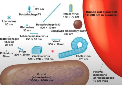
Ultramicroscopic size: Range from 20 nm to 450 nm, requiring electron microscopy for detection.
Non-cellular structure: Composed of a protein shell (capsid) surrounding a nucleic acid core.
Nucleic acid: Either DNA or RNA, never both; can be single or double-stranded.
Inactive outside host: Viruses are inert macromolecules outside the host cell and become active only inside.
Replication: Viruses hijack the host cell's genetic machinery to synthesize and assemble new virions.
Lack metabolic enzymes: Cannot synthesize proteins or generate ATP independently.
Virion
A virion is a fully formed virus capable of establishing an infection in a host.
Viral Classification
Criteria for Classification
Viruses are classified based on:
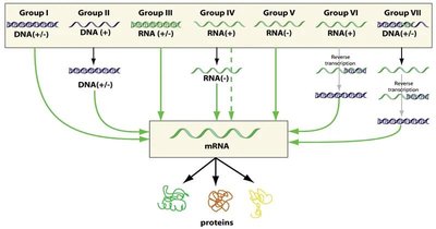
Type of nucleic acid: DNA or RNA, single or double-stranded.
Morphology: Shape and structure of the capsid and presence or absence of an envelope.
Replication methods: How the virus replicates its genome and assembles new virions.
Structure of Viruses
Basic Components
The structure of a virus can be divided into two main parts:
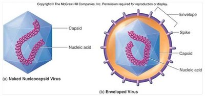
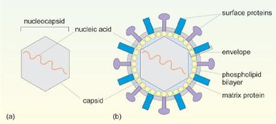
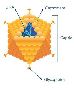
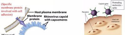
Covering: All viruses have a capsid (protein shell); some have an envelope (membrane) derived from the host cell.
Central Core: Contains the nucleic acid (DNA or RNA); some viruses also have matrix proteins and enzymes.
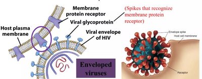
Naked vs. Enveloped Viruses
Naked viruses: Lack an envelope, contain only capsid and nucleic acid, and are more resistant to antimicrobial chemicals.
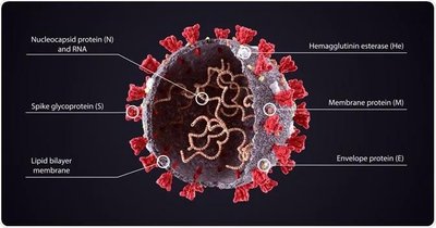
Enveloped viruses: Have a phospholipid membrane envelope derived from the host cell; more sensitive to lipid-soluble antimicrobial chemicals.
Spikes: Glycoprotein projections on the envelope essential for attachment to host cells.
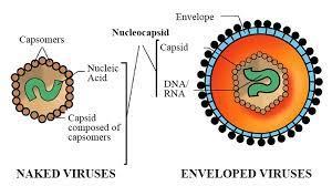
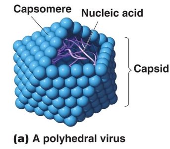
Capsid and Capsomeres
Capsid: Protein coat that encloses and protects the viral nucleic acid.
Capsomeres: Identical protein subunits that make up the capsid.
Envelope
Envelope: Found in some viruses, surrounds the capsid, composed of host cell membrane.
Spikes: Carbohydrate-protein complexes essential for attachment to host cells.
Pleomorphism: Enveloped viruses can have variable shapes due to the fluid nature of the membrane.
Function of Capsid/Envelope
Protects the nucleic acid when the virion is outside the host cell.
Helps bind the virion to a cell surface and assists penetration of viral DNA or RNA into a suitable host cell.
Viral Enzymes
Some viruses contain enzymes necessary for their replication:
Polymerases: Synthesize new strands of DNA or RNA.
Replicase: Copies RNA.
Reverse transcriptase: Synthesizes DNA from RNA (e.g., HIV).
Lysozyme: Degrades bacterial cell walls (found in bacteriophages).

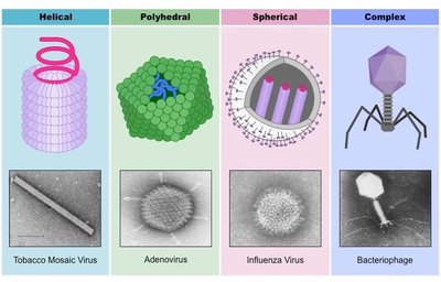
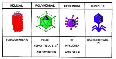
Morphology of Viruses
Types of Viral Shapes
Viruses exhibit four main morphological types:
Helical: Hollow cylinder with nucleic acids inside (e.g., Ebola, Tobacco mosaic virus).
Polyhedral: Many-sided, often icosahedral (e.g., adenovirus).
Complex: Bacteriophage with icosahedral head and helical tail sheath.
Enveloped: Roughly spherical but pleomorphic (e.g., influenza, HIV).
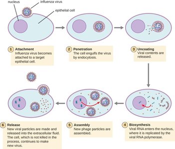
Multiplication of Animal Viruses
General Phases
The multiplication cycle of animal viruses consists of several distinct phases:
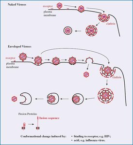
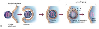
Adsorption (Attachment): Virus binds to specific molecules on the host cell surface.
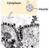
Penetration: Genome enters the host cell, often via receptor-mediated endocytosis or fusion.
Uncoating: Viral nucleic acid is released from the capsid.
Synthesis: Viral components are produced using host cell machinery.
Assembly: New viral particles are constructed.
Release: Viruses are released by budding (exocytosis) or cell lysis.
Adsorption and Host Range
Attachment occurs via specific receptors on the host cell membrane.
Host range is determined by the availability of appropriate attachment sites.
Viruses can have restricted, intermediate, or broad host ranges.
Penetration and Uncoating
Enveloped viruses: Enter via endocytosis or direct fusion with the host cell membrane.
Naked viruses: Enter via receptor-mediated endocytosis, direct penetration, or endosomal escape.
Uncoating: Release of viral nucleic acid into the cytoplasm, facilitated by host or viral enzymes.


Synthesis and Maturation
DNA viruses replicate in the nucleus; RNA viruses replicate in the cytoplasm.
Positive-sense RNA viruses can be directly translated; negative-sense RNA viruses require conversion to mRNA.
Maturation involves assembly of viral components into complete virions.
Mechanisms of Release
Budding: Enveloped viruses exit the cell by budding, acquiring a portion of the host membrane.
Lysis: Non-enveloped and complex viruses are released when the host cell ruptures.
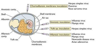
Growing Animal Viruses in the Laboratory
Methods
Injection into living animals.
Inoculation into embryonated eggs.
Cell cultures: Detection via cytopathic effects (CPE).
Cytopathic Effects and Viral-Induced Cancer
Cytopathic Effects (CPE)
Changes in cell size and shape.
Cytoplasmic and nuclear inclusion bodies.
Cell fusion, lysis, DNA alterations, and transformation into cancerous cells.
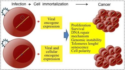
Viral-Induced Cancer
Some viruses permanently alter host genetic material, leading to cancer (oncoviruses).
Examples: Papillomavirus (cervical cancer), Epstein-Barr virus (Burkitt’s lymphoma).
Approximately 10% of cancers are virally induced.
Antiviral Drugs and Interferons
Modes of Action
Entry inhibitors: Block attachment, fusion, and uncoating (e.g., Tamiflu).
Nucleic acid synthesis inhibitors: Inhibit viral polymerase, reverse transcriptase, integrase (e.g., Acyclovir, AZT).
Assembly and release inhibitors: Target protease enzymes (e.g., Saquinovir, Tamiflu).
Interferons
Produced naturally by virally infected cells; signal neighboring cells to inhibit viral replication.
Used therapeutically to enhance immune response and inhibit cancer cells.
Types of Viral Infections
Acute, Latent, and Persistent Infections
Acute infection: Rapid onset, brief symptoms, resolution within days.
Latent infection: Virus remains dormant in host cells, may reactivate (e.g., herpes virus).
Persistent infection: Gradual increase in virions over time, often fatal (e.g., measles, HPV).
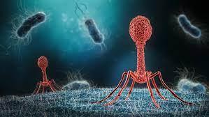
Bacteriophages
Structure and Multiplication
Bacteriophages are viruses that infect bacteria, exhibiting unique structural and replication features.
Only the nucleic acid enters the bacterial cytoplasm; no uncoating required.
Release occurs via cell lysis, not budding.
Bacteriophages are grown in bacteria and detected by plaque formation.
Lytic Cycle
The lytic cycle consists of:
Attachment (adsorption): Phage attaches to host cell via tail fibers.
Penetration: Lysozyme degrades cell wall; DNA injected into cell.
Biosynthesis: Production of phage DNA and proteins.
Maturation: Assembly of phage particles.
Release: Cell wall is broken, phages released.
Lysogenic Cycle
Phage DNA incorporates into host DNA as a prophage.
Host cell replicates prophage DNA; can later enter lytic cycle.
Lysogenic conversion can confer new properties, such as toxin production.
Prions, Viroids, and Satellite Viruses
Prions
Proteinaceous infectious particles; misfolded proteins with no nucleic acid.
Cause fatal neurodegenerative diseases (e.g., scrapie, BSE, CJS).
Extremely resistant to sterilization techniques.
Viroids and Satellite Viruses
Viroids: Short pieces of RNA without a protein coat; infect plants.
Satellite viruses: Require other viruses for replication (e.g., adeno-associated virus).
Additional info: Academic context and explanations have been expanded for clarity and completeness.
