 Back
BackViruses and Prions: Structure, Classification, and Replication
Study Guide - Smart Notes
 Tailored notes based on your materials, expanded with key definitions, examples, and context.
Viruses: General Characteristics
Definition and Properties
Viruses are minuscule, acellular infectious agents that possess unique properties distinguishing them from cellular life forms. They are obligate intracellular parasites, meaning they require living host cells to multiply.
Obligatory intracellular parasites: Viruses cannot reproduce or carry out metabolic processes independently; they must hijack a host cell's machinery.
Genetic material: Viruses contain either DNA or RNA, but never both. Their genomes may be single- or double-stranded, linear or circular, and are much smaller than those of cells.
Protein coat: The viral genome is surrounded by a protein coat called a capsid.
No ribosomes or ATP-generating mechanisms: Viruses lack the cellular machinery for protein synthesis and energy production.
Extracellular and intracellular states: Outside a cell, a virus exists as a virion (nucleic acid + capsid, sometimes with an envelope). Inside a cell, the capsid is removed, and the virus exists as nucleic acid.
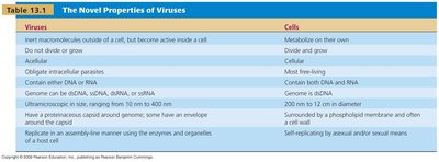
Comparison of Viruses and Cells
Viruses differ fundamentally from cells in their structure and function.
Property | Viruses | Cells |
|---|---|---|
Metabolism | Inactive outside cell | Active |
Growth | No | Yes |
Cellular structure | Acellular | Cellular |
Genetic material | DNA or RNA | DNA (and RNA) |
Protein coat | Capsid | Membrane |
Replication | Assembly in host | Self-replicating |
Structure and Morphology of Viruses
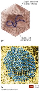
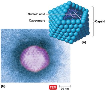
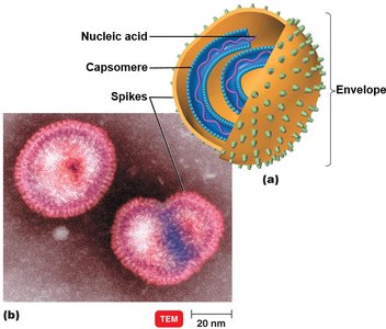
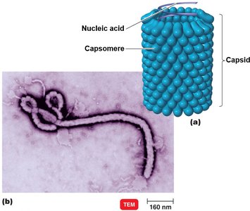
Virion Structure
The complete, fully developed viral particle is called a virion.
Nucleic acid: DNA or RNA, single- or double-stranded, linear or circular.
Capsid: Protein coat made of subunits called capsomeres.
Envelope: Some viruses have a lipid, protein, and carbohydrate envelope surrounding the capsid.
Spikes: Projections from the envelope or capsid, important for attachment to host cells.
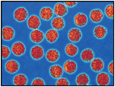
Viral Morphology
Viruses are classified by their shape and structure:
Helical viruses: Hollow, cylindrical capsid.
Polyhedral viruses: Many-sided, often icosahedral.
Enveloped viruses: Surrounded by a lipid envelope.
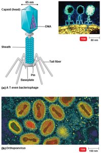
Complex viruses: Complicated structures, such as bacteriophages.
Host Range and Viral Size
Host Range
The host range of a virus is determined by specific host attachment sites and cellular factors. Most viruses infect only specific types of cells in one host, but some are generalists.
Bacteriophages: Viruses that infect bacteria.
All types of organisms: Susceptible to some virus.
Viral Size
Viruses range from 20 nm to 1000 nm in length, much smaller than most cells. 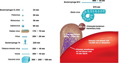
Taxonomy and Classification of Viruses
Viral Taxonomy
Genus names: End in -virus.
Family names: End in -viridae.
Viral species: Group of viruses sharing genetic information and ecological niche (host).
Subspecies: Designated by a number.
No binomial nomenclature: Unlike bacteria.
Isolation, Cultivation, and Identification of Viruses
Growing Bacteriophages
Bacteriophages are grown in bacteria, forming plaques (clearings) on a lawn of bacteria on agar. Each plaque corresponds to a single virus and can be expressed as plaque-forming units (PFU). 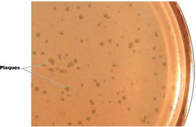
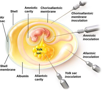
Growing Animal Viruses
Animal viruses can be grown in living animals, embryonated eggs, or cell cultures.
Embryonated eggs: Virus injected into the egg; viral growth is signaled by changes or death of the embryo.
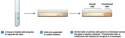
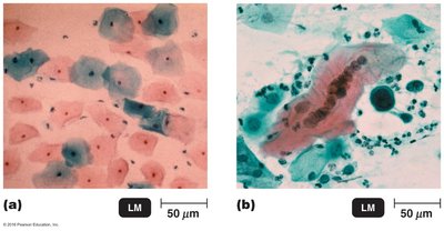
Cell cultures: Tissues are treated with enzymes to separate cells; virally infected cells are detected via cytopathic effect (CPE); continuous cell lines are used.
Viral Identification
Cytopathic effects: Observable changes in host cells.
Serological tests: Reaction of virus with antibodies.
Nucleic acids: PCR and other molecular techniques.
Viral Replication and Multiplication
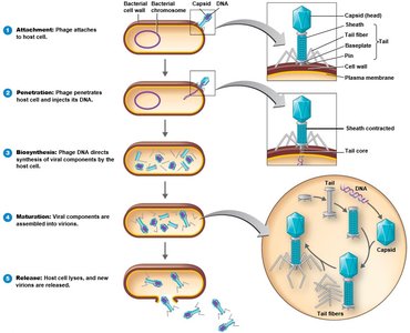
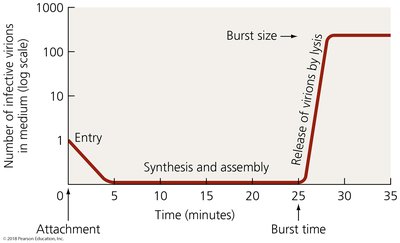
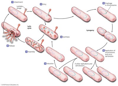
Lytic Cycle of Bacteriophages
The lytic cycle results in lysis and death of the host cell.
Attachment: Phage attaches by tail fibers to host cell.
Penetration: Phage lysozyme opens cell wall; tail sheath contracts to inject DNA.
Biosynthesis: Production of phage DNA and proteins.
Maturation: Assembly of phage particles.
Release: Phage lysozyme breaks cell wall, releasing new virions.
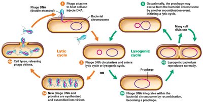
Lysogenic Cycle of Bacteriophage Lambda (λ)
In the lysogenic cycle, phage DNA incorporates into host cell DNA as a prophage, remaining latent.
Lysogeny: Phage remains latent; prophage DNA is replicated with host chromosome.
Phage conversion: Host cell exhibits new properties.
Specialized transduction: Specific bacterial genes transferred to another bacterium via a phage.
Multiplication of Animal Viruses
Attachment: Viruses attach to cell membrane.
Entry: By receptor-mediated endocytosis or fusion.
Uncoating: By viral or host enzymes.
Biosynthesis: Production of nucleic acid and proteins.
Maturation: Assembly of nucleic acid and capsid proteins.
Release: By budding (enveloped viruses) or rupture.
Biosynthesis of DNA and RNA Viruses
DNA Viruses
Replicate DNA in the nucleus using viral enzymes.
Synthesize capsid in the cytoplasm using host cell enzymes.
RNA Viruses
Multiply in host cell's cytoplasm using RNA-dependent RNA polymerase.
ssRNA (+) strand: Viral RNA serves as mRNA for protein synthesis.
ssRNA (–) strand: Viral RNA is transcribed to a + strand to serve as mRNA.
dsRNA: Double-stranded RNA viruses.
Retroviruses
Single-stranded RNA viruses that produce DNA using reverse transcriptase.
Viral DNA integrates into host chromosome as a provirus.
Examples: Lentivirus (HIV), Oncoviruses.
Viruses and Cancer
Oncogenes and Transformed Cells
Oncogenes: Genes that promote cell growth and division; uncontrolled activation can lead to cancer.
Transformed cells: Cells that have been changed by oncogenic viruses to exhibit tumor-specific antigens.
Viral Causes of Cancer
Viruses cause 20–25% of human cancers.
Some carry copies of oncogenes; some promote host oncogenes; some interfere with tumor repression.
Examples: Hodgkin’s lymphoma (EBV), Kaposi’s sarcoma (HHV-8), Cervical cancer (HPV), Liver cancer (HBV).
RNA Oncogenic Viruses
Retroviruses (e.g., HTLV-1, HTLV-2) cause adult T cell leukemia and lymphoma.
Viral RNA is transcribed to DNA, which integrates into host DNA.
Latent and Persistent Viral Infections
Latent Viral Infections
Virus remains in asymptomatic host cell for long periods; may reactivate due to changes in immunity.
Examples: Cold sores (Herpes simplex), Shingles (Varicellovirus).
Persistent Viral Infections
Occurs gradually over a long period; generally fatal.
Examples: Cervical cancer (HPV), HIV/AIDS, Liver cancer (HBV), Subacute sclerosing panencephalitis (measles virus).
Disease | Primary Effect | Causative Virus |
|---|---|---|
Cold sores | Skin and mucous membrane lesions | Herpes simplex 1 and 2 |
Leukemia | Increased white blood cell growth | HTLV-1 and -2 |
Shingles | Skin lesions | Varicellovirus (Herpesvirus) |
Cervical cancer | Increased cell growth | Human papillomavirus |
HIV/AIDS | Decreased CD4+ T cells | HIV |
Liver cancer | Increased cell growth | Hepatitis B virus |
SSPE | Mental deterioration | Measles virus |
Prions: Proteinaceous Infectious Particles
Characteristics and Diseases
Prions are infectious proteins that cause spongiform encephalopathies, characterized by large vacuoles in brain tissue and a spongy appearance.
Transmitted by ingestion, transplantation, or contact with infected tissues.
No standard treatment; prions are destroyed by incineration or autoclaving in concentrated sodium hydroxide.
Diseases: Mad cow disease (BSE), Kuru, Creutzfeldt-Jakob disease (CJD), Fatal familial insomnia, Sheep scrapie.
Prion Structure and Mechanism
Cellular PrP: Normal, functional structure with α-helices.
Prion PrP: Disease-causing form with β-pleated sheets.
Prion PrP causes cellular PrP to refold into prion PrP, propagating the disease.
Combating Prions
Normal sterilization procedures do not deactivate prions.
Prions destroyed by incineration or autoclaving in concentrated sodium hydroxide.
Enzymes approved for prion removal from medical equipment.
Summary Table: Viruses, Viroids, Prions, and Bacterial Cells
Additional info:
Esther Lederberg discovered bacteriophage λ, the fertility (F) factor, and invented replica plating, contributing to microbial genetics.
