 Back
BackChapter 13
Study Guide - Smart Notes
 Tailored notes based on your materials, expanded with key definitions, examples, and context.
Viruses and Prions
Distinctive Features of Viruses
Viruses are unique infectious agents that differ fundamentally from cellular organisms. They are obligatory intracellular parasites, meaning they require living host cells to multiply. - Contain DNA or RNA: Viral genomes consist of either DNA or RNA, but never both. - Protein coat: All viruses have a protein coat called a capsid. - No ribosomes or ATP-generating mechanisms: Viruses rely entirely on host cell machinery for protein synthesis and energy. - Host cell dependency: Viruses multiply only within living host cells, using the host’s synthesizing machinery.
Virus Sizes
Viruses are much smaller than most bacteria and eukaryotic cells, typically ranging from 20 nm to 900 nm. 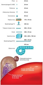
Host Range
The host range of a virus refers to the spectrum of host cells it can infect. - Specificity: Most viruses infect only specific types of cells in one host, determined by host attachment sites and cellular factors. - Bacteriophages: Viruses that infect bacteria; receptor sites may be part of the cell wall, fimbriae, or flagella. - Animal viruses: Receptor sites are typically on the plasma membrane.
Viral Structure
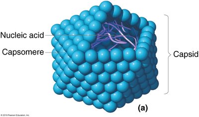
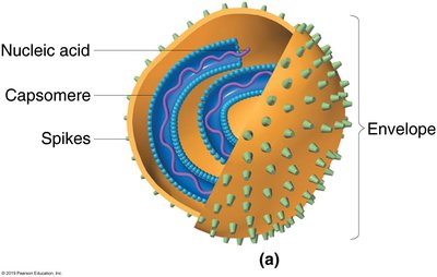
The complete, fully developed viral particle is called a virion. - Nucleic acid: DNA or RNA, single- or double-stranded, linear, circular, or segmented. - Capsid: Protein coat made of capsomeres (subunits). - Envelope: Lipid, protein, and carbohydrate coating present in some viruses. - Spikes: Projections from the outer surface, often used for attachment. 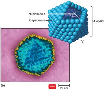
Capsid and Envelope
- Capsid: Composed of capsomeres, protects viral nucleic acid. - Envelope: Present in some viruses, derived from host cell membrane during budding. - Spikes: Used for attachment, composed of carbohydrate and protein. 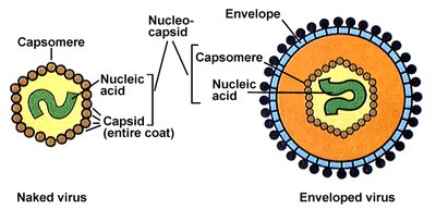
General Morphology of Viruses
Viruses exhibit several morphological types: - Helical viruses: Hollow, cylindrical capsid; e.g., rabies and Ebola virus. 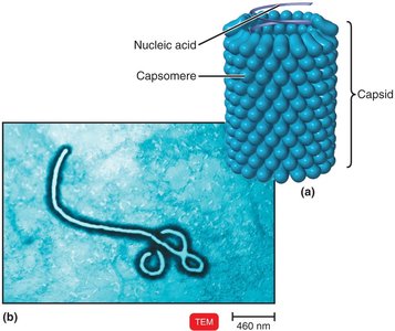 - Polyhedral viruses: Many-sided, most are icosahedral; e.g., adenoviruses and poliovirus.
  - Enveloped viruses: Roughly spherical, surrounded by an envelope.
  - Complex viruses: Complicated structures, such as bacteriophages.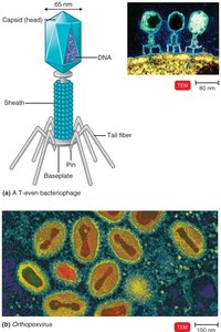
Taxonomy of Viruses
Viruses are classified based on their nucleic acid and how their mRNA is produced (Baltimore classification system). - Genus: Names end in -virus. - Family: Names end in -viridae. - Order: Names end in -ales. - Viral species: Group sharing genetic information and ecological niche.
Isolation, Cultivation, and Identification of Viruses
Bacteriophage Cultivation
- Bacteriophages are grown in bacteria and form plaques (clearings) on a lawn of bacteria on agar. - Each plaque corresponds to a single virus and can be quantified as plaque-forming units (PFU). 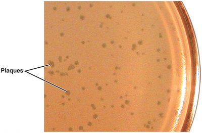
Growing Animal Viruses
- In living animals: Mice, rabbits, guinea pigs. - In embryonated eggs: Virus injected into the egg; growth signaled by changes or death of embryo. Used for vaccine production. 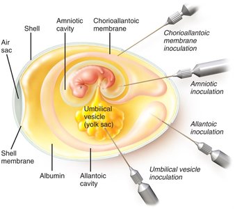 - In cell cultures: Primary cell lines (preferred), continuous cell lines (e.g., HeLa). - Cytopathic effect (CPE): Visible changes or deterioration of monolayer cells indicate viral infection.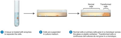
Viral Multiplication
One-Step Growth Curve
Viral multiplication involves invasion and takeover of host cell machinery. A single virion can produce thousands of progeny. The one-step growth curve illustrates the phases of viral replication. 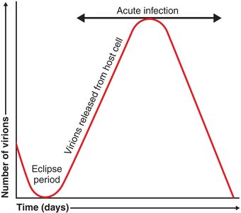
Multiplication of Bacteriophages
Bacteriophages multiply via two main mechanisms: lytic cycle and lysogenic cycle.
Lytic Cycle
- Attachment: Phage attaches to host cell. - Penetration: Phage injects DNA. - Biosynthesis: Production of phage DNA and proteins. - Maturation: Assembly of phage particles. - Release: Host cell lyses, releasing new phages. 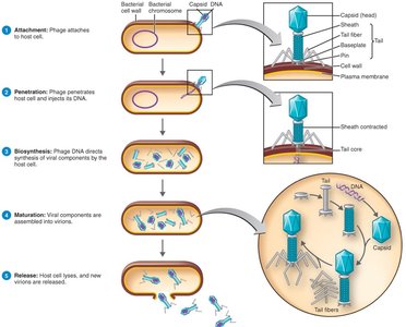
Lysogenic Cycle
- Lysogeny: Phage DNA incorporates into host DNA as a prophage. - Induction: Environmental factors may trigger the prophage to enter the lytic cycle. 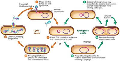 Outcomes of Lysogeny: 1. Immunity to reinfection by same phage 2. Phage conversion (new properties, e.g., toxin production) 3. Specialized transduction (transfer of specific bacterial genes)
Multiplication of Animal Viruses
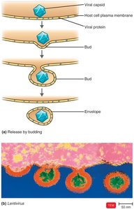
Animal viruses follow a similar but distinct process: - Attachment: Virus attaches to cell membrane. - Entry: By receptor-mediated endocytosis or fusion. - Uncoating: Viral nucleic acid separated from capsid. - Biosynthesis: Production of nucleic acid and proteins. - Maturation: Assembly of viral components. - Release: By budding (enveloped viruses) or rupture (naked viruses). 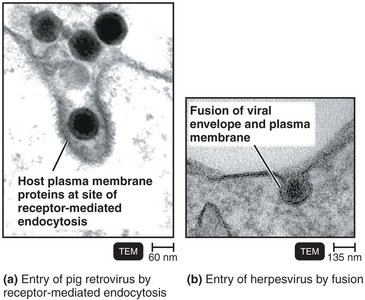
Biosynthesis of DNA Viruses
- DNA viruses replicate their DNA in the nucleus using host enzymes. - Capsid proteins are synthesized in the cytoplasm and migrate to the nucleus for assembly. 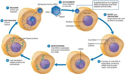
Biosynthesis of RNA Viruses That Use DNA
- Retroviruses use reverse transcriptase to produce DNA from their RNA genome. - Viral DNA integrates into the host chromosome as a provirus. - Example: Retroviridae (HIV, Lentivirus, Oncoviruses). 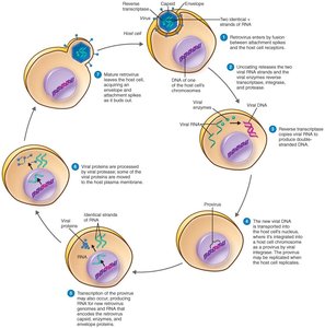
Bacteriophage and Animal Viral Multiplication Compared
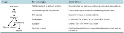
Stage | Bacteriophages | Animal Viruses |
|---|---|---|
Attachment | Tail fibers attach to cell wall proteins | Receptor sites are plasma membrane proteins and glycoproteins |
Entry | Viral DNA is injected into host cell | Capsid enters by receptor-mediated endocytosis or fusion |
Uncoating | Not required | Enzymatic removal of capsid proteins |
Biosynthesis | In cytoplasm | In nucleus (DNA viruses) or cytoplasm (RNA viruses) |
Chronic infection | Lysogeny | Latency; slow viral infections; cancer |
Release | Host cell is lysed | Enveloped viruses bud out; nonenveloped viruses rupture plasma membrane |
Viruses and Cancer
Oncogenic Viruses
Some viruses can cause cancer by integrating their genetic material into the host cell’s DNA, leading to transformation. - Proto-oncogenes: Genes that stimulate normal cell growth; mutations can convert them to oncogenes. - Oncogenes: Transform normal cells into cancerous cells. - Oncogenic viruses: 10% of cancers are virus-induced. - Examples: Epstein-Barr virus (Burkitt’s lymphoma), Human papillomavirus (HPV; cervical and anal cancer), Hepatitis B virus.
Latent and Persistent Viral Infections
Latent Viral Infections
- Virus remains in asymptomatic host cell for long periods. - All herpesviruses are capable of latency; may reactivate due to changes in immunity. - Examples: HHV1 (cold sores), Varicellovirus (shingles).
Persistent Viral Infections
- Occur gradually over a long period; generally fatal. - Example: HIV/AIDS. 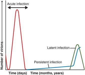
Prions
Characteristics of Prions
Prions are proteinaceous infectious particles that cause neurodegenerative diseases. - Transmission: Inherited, transmissible by ingestion, transplant, and surgical instruments. - Diseases: Spongiform encephalopathies (e.g., mad cow disease, Creutzfeldt-Jakob disease, sheep scrapie).
Prion Pathogenesis
- PrPc: Normal cellular prion protein, found on cell surface. - PrPSc: Infectious misfolded form; accumulates in brain cells, forming plaques. - Disease is caused by conversion of PrPc into PrPSc. 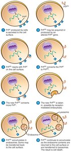 Additional info: Prion diseases are unique in that they do not involve nucleic acids, and their pathogenesis is based on protein misfolding and aggregation. ----------------------------------------
