 Back
BackViruses and Prions: Structure, Classification, Replication, and Clinical Principles
Study Guide - Smart Notes
 Tailored notes based on your materials, expanded with key definitions, examples, and context.
Viruses and Prions
General Virus Characteristics
Viruses are submicroscopic, infectious agents that are classified as nonliving microbes. They are obligate intracellular pathogens, meaning they require a host cell to replicate. Virology is the study of viruses, and understanding viruses requires foundational knowledge in cellular biology.
Size: Extremely small (30–1000 nm), acellular, and filterable.
Host Range: About 270 viruses infect humans, with many more uncharacterized.
Structure: Composed of a protein capsid and nucleic acid (DNA or RNA).
Replication: Hijack host cell machinery for replication.
Metabolism: Do not exhibit metabolism.
Characteristic | Viruses | Prokaryotes | Eukaryotes |
|---|---|---|---|
Cells? | No | Yes | Yes |
Considered alive? | No | Yes | Yes |
Relative size | Smaller than prokaryotes | Bigger than viruses, smaller than eukaryotes | Bigger than prokaryotes and viruses |
Structure | Protein capsid & nucleic acid | Cells without nuclei | Cells with nuclei |
Replication | Host cell machinery | Binary fission | Mitosis/Meiosis |
Metabolism | No | Yes | Yes |
Genome | DNA or RNA | DNA | DNA |
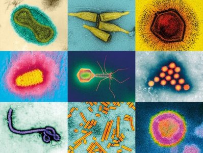
Viral Structure
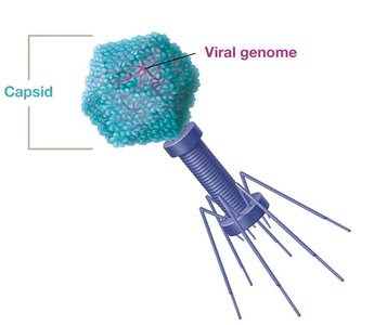
Viruses exhibit diverse structural and genomic features. The infectious virus particle is called a virion, which consists of an exterior protective protein capsid and genetic material.
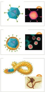
Capsid: Protein shell made of capsomere subunits; protects the genome.
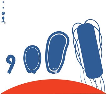
Capsid Shapes: Helical (hollow tube), icosahedral (polygon), or complex.

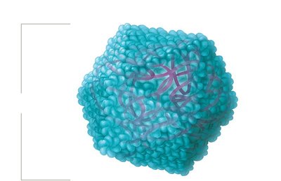
Capsid protein shells are held together by hydrophobic binding, van der Waals forces, and salt bridges at specific binding domains.
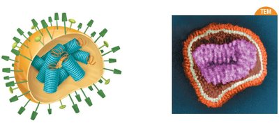
Viral Envelopes and Spikes
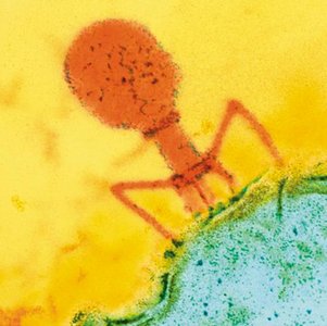
Some viruses have a lipid-based envelope derived from the host cell membrane, while others are naked (non-enveloped). Animal viruses may be enveloped or naked; bacteriophages are always naked.
Spikes (Peplomers): Glycoprotein extensions that facilitate attachment and entry into host cells; bind to specific host cell factors.
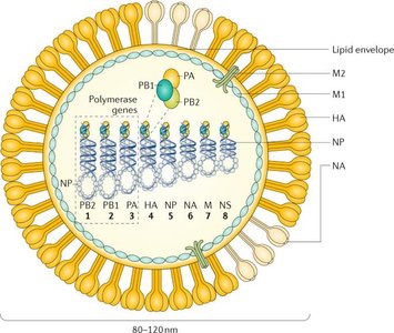
Influenza viruses mutate frequently, causing changes in spike proteins (Hemagglutinin [HA] and Neuraminidase [NA]), which define influenza subtypes (e.g., H1N1).
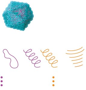
Viral Genomes
Viral genomes are highly variable and encode capsomere proteins, enzymes for replication, and structural factors. They can be DNA or RNA, single or double stranded, segmented or non-segmented, circular or linear.
DNA Viruses: Often double stranded, may be single stranded, circular or linear.
RNA Viruses: Often single stranded, may be double stranded, linear or segmented.
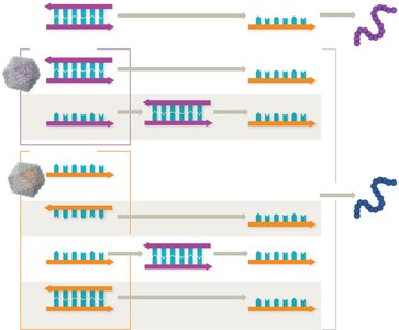
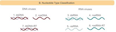
Making mRNA from Viral Genomes
Viruses must hijack host cell machinery to make viral proteins. The process of making mRNA varies by genome type:
dsDNA viruses: Transcribed by host RNA polymerases.
ssDNA viruses: Converted to dsDNA before transcription.
ssRNA (+): Genome acts as mRNA, directly translated.
ssRNA (–): Complementary to mRNA, transcribed by viral RNA-dependent RNA polymerases.
Retroviruses: RNA genome converted to DNA by reverse transcriptase, then integrated and transcribed.
dsRNA viruses: Transcribed to make mRNA by viral RNA-dependent RNA polymerases.

Viral Genome Evolution
Viruses change rapidly due to quick replication, large virion production, and high mutation rates (especially RNA viruses, which lack proofreading). Mutations can be neutral, beneficial, or deleterious.
Attenuated Strains: Reduced virulence, used in vaccines.
Reassortment: Occurs when two viral strains co-infect a cell, leading to new strains.

Antigenic Drift and Shift
Influenza viruses undergo antigenic drift (minor changes in HA/NA spikes due to mutations) and antigenic shift (major genetic reassortment, often leading to pandemics).
Antigenic Drift: Minor changes, host may lose immunity.
Antigenic Shift: Major changes, new highly virulent strains, no residual immunity.
Classifying and Naming Viruses
Classification Criteria
Viruses are classified by the International Committee on Taxonomy of Viruses (ICTV) based on:
Type of nucleic acid (DNA or RNA)
Capsid symmetry (helical, icosahedral, complex)
Envelope presence
Genome architecture (ssDNA, ssRNA, etc.)
Medically Important Virus Families
Examples of DNA and RNA virus families relevant to human health:
Family | Genome | Capsid | Envelope | Example |
|---|---|---|---|---|
Papillomaviridae | dsDNA circular | Icosahedral | Naked | HPV |
Herpesviridae | dsDNA linear | Icosahedral | Enveloped | HSV, VZV |
Poxyviridae | dsDNA linear | Complex | Enveloped | Sm. pox virus |
Flaviviridae | ssRNA+ Non-RT | Icosahedral | Enveloped | HEP C |
Retroviridae | ssRNA+ RT | Icosahedral | Enveloped | HIV |
Coronavirdae | ssRNA+ | Helical | Enveloped | COVID-19 |
Paramyxovirdae | ssRNA- | Helical | Enveloped | Measles & Mumps |
Orthomyxoviridae | ssRNA– segmented | Helical | Enveloped | Influenza |
Host Range and Tropism
Host range is the collection of species a virus can infect; tropism is the tissue or cell specificity. Some viruses have broad tropism (Ebola), others have narrow tropism (HIV).
Virus Sizes
Viruses range from 30 nm (rhinovirus, poliovirus) to 1,500 nm (pithovirus). Pathogenic animal viruses are typically 30–1,000 nm.
Naming Conventions
Viruses are named using standardized rules, with the phylum as the highest taxon. Naming conventions include order, family, subfamily, genus, and species.
Viral Replication Pathways
Generalized Steps for Animal Virus Replication
Animal virus replication involves six steps:
Attachment: Virus binds to host cell via capsid proteins (non-enveloped) or spikes (non-enveloped & enveloped).
Penetration: Entry by endocytosis/membrane fusion (enveloped), or endocytosis (non-enveloped).
Uncoating: Capsid is digested in the endocytic vesicle/cytoplasm/nucleus, genome is released.
Replication (synthesis): Genome is replicated and viral proteins are made.
Assembly: New virions are formed.
Release: Enveloped viruses bud off; non-enveloped rupture/lyse the cell during release.
Persistent Infections
Some viruses cause persistent infections, which can be chronic (continuous release of virions and slow progression of disease) or latent (flare-ups with dormancy/latency). Examples include HIV & Provirus; integrated DNA (chronic) and herpesviruses (latent).
Herpesviridae
Human herpes virus -1 (HSV-1): cold sores
HSV-2: genital herpes
Varicella-zoster virus (HHV-3): chickenpox and shingles
Oncogenic Viruses
Oncogenic viruses (oncoviruses) can cause cancer by stimulating uncontrolled cell division or reducing cell death signals.
Virus | Genome | Integrates DNA? | Cancer Link | Mechanism |
|---|---|---|---|---|
HPV | DNA Papillomaviridae family | Yes | Cervical, oropharyngeal, and anal cancers | Uncontrolled cell division |
Human herpes virus-8 | DNA; Herpesviridae family | No | Kaposi sarcoma (skin cancer) | Uncontrolled cell division |
Epstein-Barr (HHV-4 causes mononucleosis- kissing disease | DNA; Herpesviridae family | No | B-cell and T-cell lymphomas and Hodgkin's disease | Uncontrolled cell division |
HTLV | RNA; Retroviridae family | Yes | Adult T-cell leukemia | Uncontrolled cell division |
Hepatitis B | DNA; Hepadnaviridae family | No | Liver cancer | Chronic inflammation causes host cell DNA damage and mutations |
Hepatitis C | RNA; Flaviviridae family | No | Liver cancer | Chronic inflammation causes host cell DNA damage and mutations |
Clinical Aspects of Viruses
Laboratory Propagation
Viruses are propagated in the lab using cell culture, live animal hosts, or embryonated eggs. The plaque assay is used to quantify infectious viral particles (PFU/ml).
- clear zones are the plaques
Diagnostic Methods
Specificity: the test only detects the virus(es) of interest (no false positives)
Sensitivity: the test detects very low levels of the target (no false negetives)
Detection of viruses relies on molecular methods due to their small size. Key methods include:
Agglutination Tests: Use antibodies or antigens linked to beads; positive result is agglutination (stick together).
Enzyme-linked immunosorbent Assay (ELISA): Detects viral proteins or antibodies; color change indicates binding to surface.
Nucleic Acid Detection: PCR and RT-qPCR (reads amplification RNA to DNA) detect viral genetic material with high sensitivity and specificity. (very good)
RT-qPCR results: "cycle threshold" (Ct)- number of cycles for the signal to reach the threshold where it can be seen over background signal
high concentration: low Ct number- don't need much amplification to see it
Low concentration: high Ct number- need a lot of amplification to see it
Antiviral Drugs
Antiviral drugs target various steps in viral replication but rarely cure infections. Most drugs target viral enzymes and must be selectively toxic. Vaccines are crucial for prevention.
Attachment/Penetration/Uncoating Inhibitors: Block viral entry (e.g., amantadine, maraviroc).
Replication Inhibitors: Nucleoside analogs (e.g., acyclovir (DNA), ribavirin (RNA)) block nucleic acid synthesis. Stop polymerase.
Nucleoside reverse transcriptase inhibitors (NTRIs): target reverse transcriptase enzymes (e.g., Azidothymidine (AZT)).
Interferons: Naturally occurring. Signal cells to make defensive changes; can be administered therapeutically.
Release Inhibitors: Oseltamivir and zanamivir prevent influenza virion release.
Prions
Prion Structure and Diseases
Prions are infectious proteins with no genetic material; they do not replicate. They cause transmissible spongiform encephalopathies (TSEs), such as Creutzfeldt-Jakob disease and fatal familial insomnia.
Gerstmann-Straussler-Schienker Syndrome- fatal familial insomnia inherited.
Creuzfeldt-Jakob Disease (CJD)- acquired from cows
Prion-like Disorders: Neurodegenerative diseases (Alzheimer's, Parkinson's, ALS) are associated with misfolded proteins.
