 Back
BackChapter 6 Study guide
Study Guide - Smart Notes
 Tailored notes based on your materials, expanded with key definitions, examples, and context.
Viruses and Prions
General Virus Characteristics
Viruses are unique infectious agents that differ fundamentally from prokaryotic and eukaryotic cells. They are considered nonliving because they lack cellular structure and metabolic processes, and require a host cell for replication.
Viruses are submicroscopic, acellular, and obligate intracellular pathogens.
Virology is the study of viruses.
Over 5,000 mammal-infecting viral species have been described, with many more uncharacterized.
Comparison of Viruses, Prokaryotes, and Eukaryotes
Viruses differ from prokaryotes and eukaryotes in size, structure, and replication.
Viruses: 20–400 nm, acellular, require host cell for replication.
Prokaryotes: 0.5–2 µm, cellular, independent replication.
Eukaryotes: 10–100 µm, cellular, independent replication.
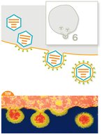
Viral Structure
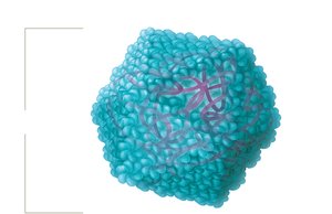
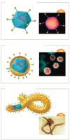
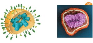
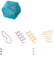
Viruses exhibit diverse structural features, including capsids, envelopes, and spikes.
Capsid: Protein shell protecting the genome, made of capsomere subunits.
Capsid shapes: Helical, icosahedral, or complex.
Envelope: Lipid-based membrane surrounding some viruses, acquired from host cell.
Spikes (Peplomers): Glycoprotein extensions aiding in host cell attachment.
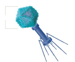
Capsid Structures
Helical capsids: Hollow tube-like structure.
Icosahedral capsids: Three-dimensional polygon.
Complex capsids: Found in bacteriophages, with additional structures for genome injection.

Viral Envelopes and Spikes
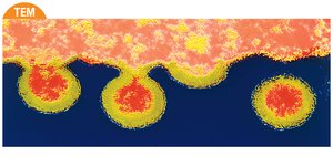
Enveloped viruses arise from budding off the host cell.
Naked viruses arise from lysing the host cell.
Bacteriophages are always naked.
Spikes bind to specific host cell factors, determining host range and tropism.
Viral Genomes
Viral genomes are highly variable and encode essential proteins for replication.
Can be DNA or RNA, single or double-stranded, segmented or nonsegmented, circular or linear.
Most viruses have fewer than 300 genes.
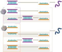
Making mRNA from Viral Genomes
The goal of all viruses is to use the host cell to make viral proteins.
dsDNA viruses: Transcribed by host RNA polymerases.
ssDNA viruses: Converted to dsDNA before transcription.
ssRNA+ viruses: Genome acts as mRNA, directly translated.
ssRNA- viruses: Genome is complementary to mRNA, transcribed by viral RNA-dependent RNA polymerases.
Retroviruses: RNA genome converted to DNA by reverse transcriptase, integrated into host genome.
dsRNA viruses: Require RNA-dependent RNA polymerases for transcription.
Viral Genome Evolution
Viruses evolve rapidly due to quick replication and high mutation rates, especially in RNA viruses.
RNA polymerases lack proofreading, leading to more mutations.
Mutations can be neutral, beneficial, or deleterious.
Reassortment occurs when two strains coinfect a cell, creating new viral strains.
Antigenic Drift and Shift
Antigenic drift: Minor changes in viral antigens due to mutations.
Antigenic shift: Major genetic reassortment, leading to new strains and potential pandemics.
Classifying and Naming Viruses
Viruses are classified by nucleic acid type, capsid symmetry, envelope presence, and genome architecture.
ICTV sets naming conventions.
Medically important families include Papillomaviridae, Herpesviridae, Picornaviridae, Orthomyxoviridae, Retroviridae, and Coronaviridae.
Host Range and Tropism
Host range: Species a virus can infect.
Tropism: Specificity for certain tissues or cell types.
Virus Sizes
Viruses range from 30 nm (rhinovirus) to 1,500 nm (pithovirus). 
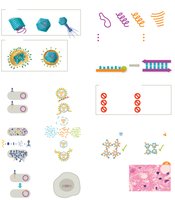
Viral Replication Pathways
Viruses hijack host cell machinery to multiply.
Bacteriophage Replication
Lytic pathway: Immediate virion production, host cell lysis.
Lysogenic pathway: Phage genome integrates into host genome (prophage), can later enter lytic cycle.
Phage conversion: Prophages confer new pathogenic properties (e.g., toxins).
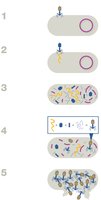
Animal Virus Replication
Six steps: Attachment, Penetration, Uncoating, Replication, Assembly, Release.
Enveloped viruses enter by fusion or endocytosis; naked viruses by endocytosis.
Release: Enveloped viruses bud; naked viruses lyse the cell.


Persistent Infections
Chronic infections: Continuous release of virions, slow disease progression (e.g., HIV).
Latent infections: Flare-ups with periods of dormancy (e.g., herpesviruses).
Oncogenic viruses: Cause cancer by stimulating uncontrolled cell division (e.g., HPV, HTLV).
Clinical Aspects of Viruses
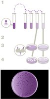
Growing Viruses in the Laboratory
Bacteriophages: Grown using plaque assays on petri plates.
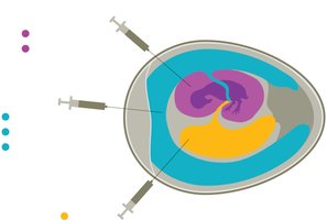
Animal viruses: Grown in tissue culture, live animals, or embryonated eggs.

Diagnostic Tests for Viral Infections
Detection methods: Molecular (PCR, sequencing), protein (ELISA, agglutination), antibody detection.
Specificity: Detects only target virus.
Sensitivity: Detects low levels of virus.

Antiviral Drugs
Antiviral drugs target various steps in viral replication but rarely cure infections.
Attachment, penetration, uncoating inhibitors (e.g., amantadine, docosanol).
Replication inhibitors: Nucleoside analogs (e.g., acyclovir, ribavirin), reverse transcriptase inhibitors (e.g., AZT).
Assembly and release inhibitors (e.g., oseltamivir, zanamivir).
Interferons: Signal cells to limit viral entry and replication.
Vaccines: Train immune system to recognize viruses.
Prions
Prions are infectious proteins that cause transmissible spongiform encephalopathies (TSEs).
No genetic material, do not replicate.
Diseases: Creuzfeldt-Jakob disease, Gerstmann-Straussler-Schienker syndrome, fatal familial insomnia.
Some neurodegenerative diseases (Alzheimer's, Parkinson's, ALS) exhibit prion-like features.
Clinical Case Study: Blood Transfusion and Viral Discovery
Case Summary
Blood transfusion in 1976 led to discovery of a new virus in cataloged samples.
Son died of liver cancer; new virus found in other transfusion recipients.
Key Questions and Concepts
Screening old blood samples: Use molecular diagnostics (PCR, sequencing).
Classification: Determine genome type, structure, host range, and tropism.
Isolation and growth: Use tissue culture or animal models.
RNA genome: Prescribe antivirals targeting RNA replication (e.g., ribavirin).
Immune reaction: Shared genes with hepatitis C virus, detected by agglutination test.
Low viral titers: Suggest chronic, low-level infection.
Oncogenic potential: Not ruled out by absence of cancer in other patients.
Summary
Viruses and prions are distinct from other microbes in structure, replication, and clinical impact. Understanding their classification, replication pathways, diagnostic methods, and treatment strategies is essential for microbiology students and clinical practice. Additional info: Expanded explanations and context were added for clarity and completeness.
