 Back
BackViruses and Prions: Structure, Replication, and Clinical Relevance
Study Guide - Smart Notes
 Tailored notes based on your materials, expanded with key definitions, examples, and context.
Viruses: Structure and Genomic Features
General Characteristics of Viruses
Viruses are minuscule, acellular, infectious agents that contain either DNA or RNA as their genetic material, but never both. They are obligate intracellular parasites, meaning they require a host cell to replicate and cannot carry out metabolic processes independently. Viruses lack cellular structures such as cytoplasmic membranes, cytosol, and organelles. They exist in two states: the extracellular state (virion) and the intracellular state (nucleic acid only).
Virion: The complete, infectious viral particle outside a host cell, consisting of a nucleic acid genome surrounded by a protein coat (capsid). Some virions possess an additional phospholipid envelope derived from the host cell membrane.
Intracellular state: The capsid is removed, and the virus exists as naked nucleic acid within the host cell.
Viral Genomes
Viral genomes are highly diverse and are a primary basis for virus classification. They may be composed of DNA or RNA, which can be single-stranded (ss) or double-stranded (ds), linear, circular, or segmented. Viral genomes are much smaller than those of cellular organisms.
DNA viruses: Often double-stranded, can be linear or circular.
RNA viruses: Often single-stranded, can be linear or segmented; some are double-stranded.
Viruses are classified based on their genome type, structure, and replication strategy.
Host Range and Tropism
The host range of a virus refers to the spectrum of host species it can infect, determined by the compatibility of viral surface proteins with host cell receptors. Tropism describes the specificity of a virus for particular tissues or cell types within a host.
Some viruses are highly specific (e.g., measles virus infects only humans), while others have a broad host range.
Tropism is dictated by viral surface factors and host cell receptors.
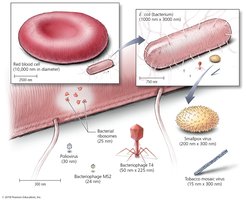
Size and Morphology of Virions
Viruses are much smaller than prokaryotic and eukaryotic cells, typically ranging from 10 to 400 nm in diameter. Their small size allows them to be visualized only by electron microscopy.
Viral Structure: Capsids and Envelopes
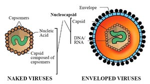
Capsid Morphology
The capsid is a protein shell composed of subunits called capsomeres. It protects the viral genome and facilitates attachment to host cells. Viruses are classified by capsid shape:
Helical: Capsomeres arranged in a spiral around the nucleic acid.
Polyhedral (Icosahedral): Capsid forms a roughly spherical shape with 20 triangular faces.
Complex: Capsid structures that do not fit into the other categories, often seen in bacteriophages.

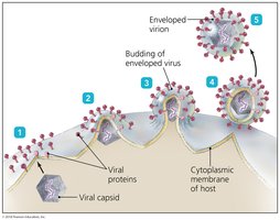
Viral Envelope
Some animal viruses possess an envelope derived from the host cell membrane during viral replication or release. The envelope contains viral glycoproteins (spikes) that facilitate host cell recognition and entry. Enveloped viruses are generally more sensitive to environmental conditions than naked viruses.
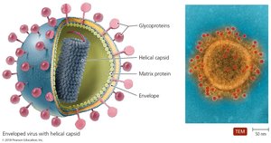
Viral Spikes (Peplomers)
Spikes are glycoprotein extensions that protrude from the capsid or envelope, mediating attachment and entry into host cells. They are highly specific for host cell receptors and are targets for immune responses and antiviral drugs.
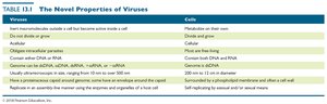
Comparison of Viruses and Cells
The following table summarizes the novel properties of viruses compared to cells:
Viruses | Cells |
|---|---|
Obligate intracellular parasites | Metabolize and grow independently |
Contain either DNA or RNA, never both | Contain both DNA and RNA |
Acellular | Cellular |
Do not divide or grow | Divide and grow |
Replicate by assembly of subunits | Self-replicating by binary fission or mitosis |
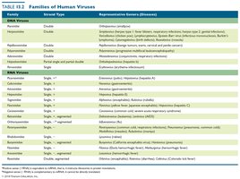
Classification of Viruses
Families of Human Viruses
Viruses are classified into families based on genome type, capsid symmetry, presence or absence of an envelope, and replication strategy. The following table lists major families of human viruses and their representative diseases:
Viral Replication Cycles
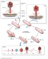
Lytic Replication Cycle
The lytic cycle is a replication process that results in the destruction (lysis) of the host cell and release of new virions. The five stages are:
Attachment: Virus binds to host cell surface.
Entry: Viral genome enters the host cell.
Synthesis: Host machinery synthesizes viral components.
Assembly: New virions are assembled.
Release: Host cell lyses, releasing new virions.
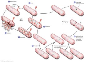
Lysogenic Replication Cycle
Some bacteriophages undergo a lysogenic cycle, where the viral genome integrates into the host chromosome as a prophage and replicates along with the host cell. The prophage may later enter the lytic cycle. Lysogenic conversion can confer new properties (e.g., toxin production) to the host bacterium.
Replication of Animal Viruses
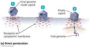
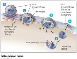
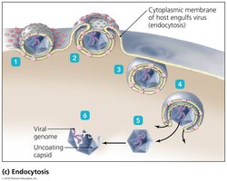
Animal viruses follow similar steps as bacteriophages but differ in entry and release mechanisms due to the presence of an envelope and the eukaryotic nature of host cells. Entry can occur via direct penetration, membrane fusion, or endocytosis.
Direct penetration: Viral genome enters the cell directly.
Membrane fusion: Viral envelope fuses with host membrane, releasing the capsid.
Endocytosis: Virus is engulfed by the host cell membrane.
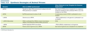
Synthesis Strategies of Animal Viruses
The synthesis of viral components depends on the type of viral genome. DNA viruses often replicate in the nucleus, while RNA viruses typically replicate in the cytoplasm. Retroviruses use reverse transcriptase to synthesize DNA from their RNA genome.
Assembly and Release
Most DNA viruses assemble in the nucleus, while most RNA viruses assemble in the cytoplasm. Enveloped viruses are released by budding, causing persistent infections, while naked viruses are released by lysis or exocytosis.
Latency and Persistent Infections
Some animal viruses can remain dormant (latent) within host cells for years. Latent viruses (proviruses) may integrate into the host genome, and their reactivation can lead to recurrent disease. Chronic infections involve continuous release of virions and slow disease progression.
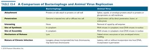
Comparison of Bacteriophage and Animal Virus Replication
Viruses and Cancer
Oncogenic Viruses
Some viruses can induce cancer by disrupting normal cell cycle regulation. They may carry oncogenes, activate host oncogenes, or interfere with tumor suppressor genes. Examples include human papillomavirus (HPV) and Epstein-Barr virus (EBV).
Other Parasitic Particles: Viroids and Prions
Viroids
Viroids are small, circular, single-stranded RNA molecules that infect plants. They lack a protein coat and do not encode proteins. Viroid RNA can bind to complementary plant RNA, leading to degradation and disease.
Prions
Prions are infectious proteins that lack nucleic acids. They cause transmissible spongiform encephalopathies (TSEs) by inducing abnormal folding of normal cellular prion protein (PrP) into a disease-causing form. Prion diseases include Creutzfeldt-Jakob disease, mad cow disease, and kuru.
Prions do not replicate; ACELLULAR
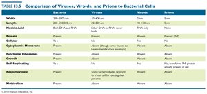
Comparison of Viruses, Viroids, Prions, and Bacteria
Case Study: Influenza Virus
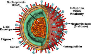
Structure and Key Features
The influenza virus is an enveloped, negative-sense, single-stranded RNA virus with a segmented genome. It possesses two major surface glycoproteins: hemagglutinin (HA) for attachment and neuraminidase (NA) for release. The segmented genome allows for genetic reassortment, contributing to antigenic variation.
Antigenic Drift and Shift
Antigenic drift refers to minor mutations in viral genes encoding HA and NA, leading to seasonal epidemics. Antigenic shift is a major genetic reassortment event that can result in new viral subtypes and pandemics, as the population lacks immunity to the novel strain.
Antiviral Drugs and Prevention
Mechanisms of Antiviral Drugs
Antiviral drugs target various stages of the viral replication cycle, including attachment, penetration, uncoating, replication, assembly, and release. Examples include:
Entry inhibitors: Block viral attachment or fusion (e.g., maraviroc, docosanol).
Nucleoside analogs: Mimic nucleotides to halt viral genome replication (e.g., acyclovir, ribavirin).
Reverse transcriptase inhibitors: Block retroviral replication (e.g., AZT for HIV).
Neuraminidase inhibitors: Block release of influenza virions (e.g., oseltamivir).
Interferons: Enhance host antiviral defenses.
Vaccination remains the most effective strategy for preventing serious viral diseases.
