 Back
BackViruses and Prions: Structure, Replication, and Clinical Relevance
Study Guide - Smart Notes
 Tailored notes based on your materials, expanded with key definitions, examples, and context.
Viruses and Prions
Introduction to Viruses
Viruses are submicroscopic, acellular infectious agents that require a host cell for replication. The study of viruses is known as virology. Viruses can infect all forms of life, including bacteria (bacteriophages), animals, and plants. They are considered nonliving because they lack cellular structure and metabolism.
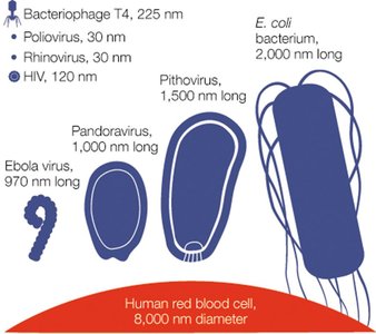
Size: Typically 20–400 nm, much smaller than prokaryotic and eukaryotic cells.
Obligate intracellular pathogens: Cannot reproduce outside a host cell.
Host range: Viruses can infect every branch in the tree of life.
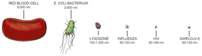
Comparison of Viruses, Prokaryotes, and Eukaryotes
Viruses differ fundamentally from prokaryotic and eukaryotic cells in structure, replication, and metabolism.
Characteristic | Viruses | Prokaryotes | Eukaryotes |
|---|---|---|---|
Cells? | No | Yes | Yes |
Considered alive? | No | Yes | Yes |
Relative size | Smaller than prokaryotes | Bigger than viruses, smaller than eukaryotes | Bigger than prokaryotes and viruses |
Structure | Protein capsid, nucleic acid | Cells without nuclei | Cells with nuclei |
Replication | Hijack host machinery | Binary fission | Mitosis/Meiosis |
Metabolism | No | Yes | Yes |
Genome | DNA or RNA | DNA | DNA |
Viral Structure and Genomic Features
Virion Structure
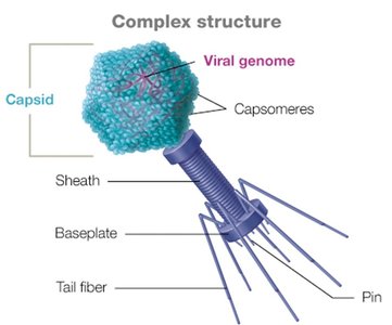
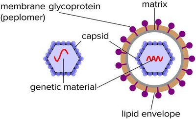
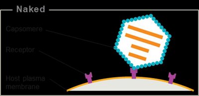
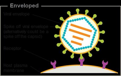
A virion is a single, infectious virus particle. It consists of a protective protein shell called a capsid, which encloses the viral genome (either DNA or RNA). Some viruses also possess an outer lipid envelope derived from the host cell membrane.
Capsid: Made of protein subunits called capsomeres.
Envelope: Lipid-based, present in some animal viruses.
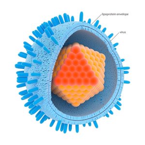
Capsid Symmetry
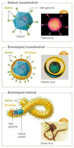
Helical capsids: Hollow tube-like structure (e.g., tobacco mosaic virus).
Icosahedral capsids: Three-dimensional polygons (e.g., adenovirus, coronavirus).
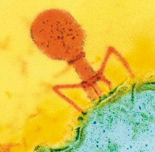
Complex capsids: More elaborate structures, often seen in bacteriophages.
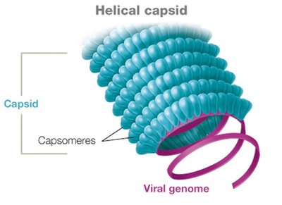
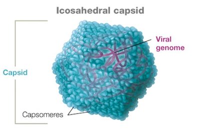
Viral Envelopes and Spikes
Enveloped viruses have a lipid membrane surrounding the capsid, acquired from the host cell during budding. Naked viruses lack this envelope and are released by cell lysis. Many viruses have surface proteins called spikes (peplomers) that facilitate attachment to host cells.
Enveloped viruses: e.g., influenza, herpes, coronaviruses.
Naked viruses: e.g., poliovirus, human papillomavirus.
Bacteriophages: Always naked, released by lysis.
Viral Genomes
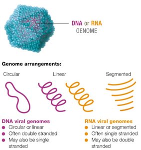
Viral genomes are highly diverse and can be composed of DNA or RNA, which may be single- or double-stranded, linear, circular, or segmented. Most viruses have fewer than 300 genes, encoding structural proteins, enzymes for replication, and other factors.
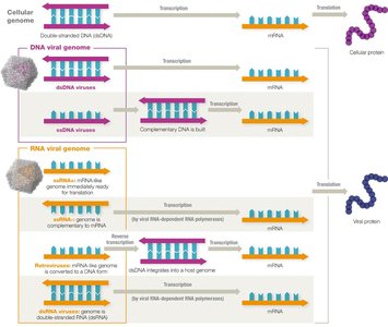
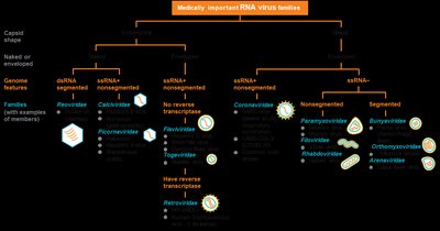
Genome types: dsDNA, ssDNA, dsRNA, ssRNA (positive or negative sense).
Segmented genomes: Genome divided into separate pieces (e.g., influenza virus).
Viral Replication and Mutation
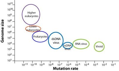
Viruses replicate rapidly and produce large numbers of progeny, especially RNA viruses, which have high mutation rates due to lack of proofreading by RNA polymerases. This leads to rapid evolution and emergence of new strains.
Mutation effects: Neutral, beneficial, or detrimental.
Attenuated strains: Weakened viruses used in vaccines.
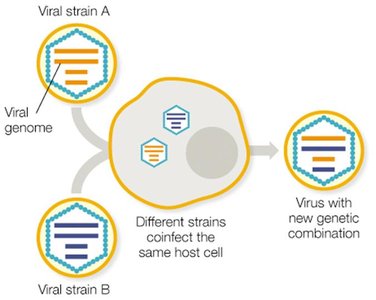
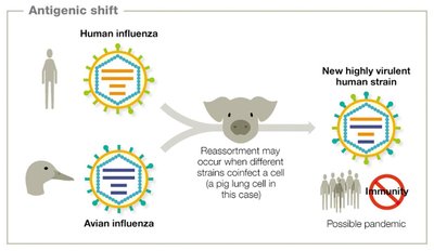
Reassortment: Exchange of genome segments between different viral strains infecting the same cell, leading to new combinations.
Antigenic Drift and Shift
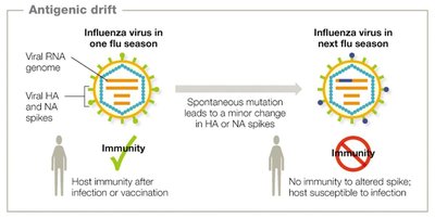
Influenza viruses are notable for frequent changes in their surface antigens (spikes), which can occur by two mechanisms:
Antigenic drift: Minor changes due to point mutations in HA and NA spikes, leading to seasonal epidemics.
Antigenic shift: Major genetic reassortment, often resulting in new, highly infectious strains and pandemics.
Classification and Naming of Viruses
Classification Criteria
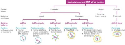
Viruses are classified based on:
Type of nucleic acid (DNA or RNA)
Capsid symmetry (helical, icosahedral, complex)
Presence or absence of an envelope
Genome architecture (e.g., ssDNA, dsRNA)
Naming Conventions
Taxon | Example | Notes |
|---|---|---|
Order | Herpesvirales | Ends in 'virales' |
Family | Herpesviridae | Ends in 'viridae' |
Subfamily | Alphaherpesvirinae | Ends in 'virinae' |
Genus | Simplexvirus | Ends in 'virus' |
Species | Human herpesvirus-1 | Not abbreviated |
Host Range and Tropism
Host Range
The host range of a virus is the spectrum of species it can infect. Some viruses are species-specific, while others can infect multiple species due to genetic changes or reassortment.
Example: Measles virus infects only humans; avian influenza can infect birds and, after reassortment, humans.
Tropism
Tropism refers to the specificity of a virus for particular host tissues or cell types, determined by viral surface proteins and host cell receptors.
Broad tropism: Ebola virus infects many cell types.
Narrow tropism: Hepatitis viruses infect primarily liver cells.
Viral Replication
Bacteriophage Replication
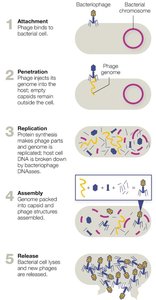
Bacteriophages infect bacteria and replicate via two main pathways: lytic and lysogenic cycles.
Lytic cycle: Virus immediately replicates, lyses host cell, and releases new virions.
Lysogenic cycle: Viral genome integrates into host DNA as a prophage, replicates with host, can later enter lytic cycle.
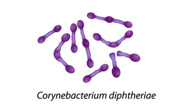
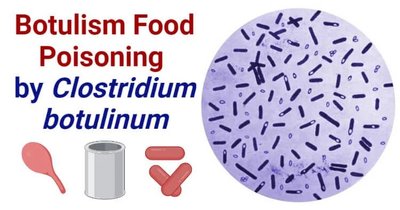
Phage conversion: Prophages can confer new pathogenic traits to bacteria (e.g., toxin production in Corynebacterium diphtheriae and Clostridium botulinum).
Animal Virus Replication
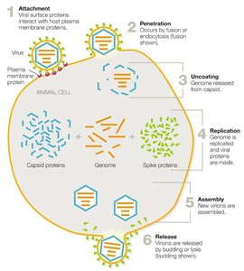
Animal viruses follow a generalized replication cycle with six main steps:
Attachment: Virus binds to host cell receptors via capsid proteins or spikes.
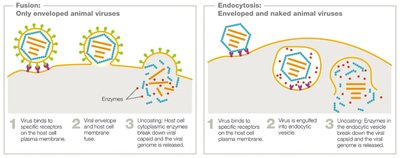
Penetration: Entry by endocytosis or membrane fusion.
Uncoating: Capsid is removed, releasing viral genome.
Replication (Synthesis): Genome is replicated, viral proteins are synthesized.
Assembly: New virions are assembled.
Release: Enveloped viruses bud off; naked viruses lyse the host cell.
Persistent Viral Infections and Oncogenesis
Types of Persistent Infections
Acute infections: Rapid onset, short duration (e.g., influenza).
Chronic infections: Continuous release of virions, slow disease progression (e.g., HIV).
Latent infections: Periods of dormancy with intermittent flare-ups (e.g., herpesviruses).
Oncogenic Viruses
Some viruses can cause cancer by integrating into the host genome or inducing chronic inflammation, leading to uncontrolled cell division.
Virus | Genome | Integrates? | Cancer Link | Mechanism |
|---|---|---|---|---|
HPV | DNA | Yes | Cervical, oropharyngeal, anal cancers | Uncontrolled cell division |
HHV-8 | DNA | No | Kaposi sarcoma | Uncontrolled cell division |
EBV | DNA | No | Lymphomas, Hodgkin’s disease | Uncontrolled cell division |
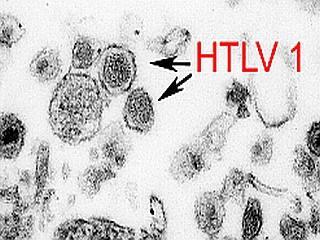
HTLV | RNA | Yes | Adult T-cell leukemia | Uncontrolled cell division |
Hepatitis B | DNA | No | Liver cancer | Chronic inflammation |
Hepatitis C | RNA | No | Liver cancer | Chronic inflammation |
Virus Detection and Cultivation
Plaque Assays
Plaque assays are used to quantify bacteriophages. Each clear zone (plaque) on a bacterial lawn represents lysis by a single phage. The number of plaque-forming units (PFUs) indicates viral titer.
Growing Animal Viruses
Animal viruses are cultivated in tissue cultures, embryonated eggs, or live animal hosts. Tissue culture is the most common method for laboratory propagation.
Diagnostic Methods
Latex agglutination tests: Detect viral antigens or antibodies using latex beads.
ELISA: Enzyme-linked immunosorbent assay for detecting viral proteins or antibodies.
Nucleic acid detection: PCR, sequencing, and fluorescent probes for viral DNA/RNA.
Key test qualities: Specificity (no false positives) and sensitivity (no false negatives).
Antiviral Drugs and Vaccines
Antiviral Drug Mechanisms
Entry inhibitors: Block viral attachment or penetration (e.g., docosanol, palivizumab).
Nucleoside analogs: Mimic nucleotides, inhibit viral genome replication (e.g., acyclovir, ribavirin).
Reverse transcriptase inhibitors: Block retroviral replication (e.g., AZT).
Antisense antivirals: Bind viral RNA, prevent translation (e.g., Vitravene).
Interferons: Signal uninfected cells to mount antiviral defenses.
Neuraminidase inhibitors: Prevent influenza virion release (e.g., oseltamivir).
Most antivirals limit, but do not cure, infections. Vaccination is crucial for prevention.
Prions
Prion Diseases
Prions are infectious proteins that lack nucleic acids. They cause transmissible spongiform encephalopathies (TSEs), which are fatal neurodegenerative diseases. Prions induce misfolding of normal proteins in the brain, leading to tissue degeneration and characteristic sponge-like holes.
Examples: Creutzfeldt-Jakob disease, mad cow disease.
Diagnosis: Detection of spongiform changes in brain tissue post-mortem.
Prion-like Mechanisms in Neurodegenerative Diseases
Some neurodegenerative diseases, such as Alzheimer's, Parkinson's, and ALS, exhibit prion-like mechanisms, where misfolded proteins propagate by inducing misfolding in normal proteins. However, these are not true prion diseases.
