 Back
BackViruses and Prions: Structure, Replication, and Pathogenesis
Study Guide - Smart Notes
 Tailored notes based on your materials, expanded with key definitions, examples, and context.
Viruses and Prions
Main Differences Between Viruses, Bacteria, and Prions
Viruses, bacteria, and prions are all infectious agents, but they differ significantly in structure, replication, and pathogenic mechanisms.
Bacteria: Prokaryotic, cellular organisms that reproduce by binary fission, possess both DNA and RNA, have ribosomes, and generate ATP through metabolism.
Viruses: Acellular entities composed of nucleic acid (DNA or RNA) surrounded by a protein coat (capsid), sometimes with a lipid envelope. They are obligate intracellular parasites and lack ribosomes and ATP-generating metabolism.
Prions: Infectious proteins lacking nucleic acids. They cause disease by inducing abnormal folding of specific normal cellular proteins.
Intracellular Pathogen: An organism or agent that must invade and replicate within a host cell to survive and reproduce.
Obligate Intracellular Pathogen: An organism or agent that cannot reproduce outside the host cell (e.g., viruses, some bacteria like Chlamydia and Rickettsia).
Comparison Table: Bacteria, Viruses, and Prions
Feature | Typical Bacteria | Intracellular Bacteria (Rickettsias/Chlamydias) | Viruses | Prions |
|---|---|---|---|---|
Intracellular Parasite/Pathogen | No | Yes | Yes | No |
Plasma Membrane | Yes | Yes | No | No |
Binary Fission | Yes | Yes | No | No |
Pass through Bacteriological Filters | No | Yes | Yes | Yes |
Possess Both DNA and RNA | Yes | Yes | No (one or the other) | No |
ATP-Generating Metabolism | Yes | No | No | No |
Ribosomes | Yes | Yes | No | No |
Sensitivity to Antibiotics | Yes | Yes | No | No |
Sensitivity to Antivirals | No | No | Yes | No |
Sensitivity to Interferon | No | No | Yes | No |
General Structure of a Virus
Virion Components
A virion is a complete, infectious virus particle. Its main components include:
Capsid: Protein coat surrounding the viral nucleic acid.
Nucleocapsid: The combination of the viral genome and the capsid.
Envelope: (In enveloped viruses) A phospholipid membrane derived from the host cell, covering the nucleocapsid.
Spikes: Glycoprotein projections from the envelope, involved in host cell recognition and attachment.
Types of Viruses
Naked (Non-enveloped) Viruses: Consist only of a capsid and genome; lack an envelope. Viral attachment proteins are capsomeres.
Enveloped Viruses: Possess a lipid envelope with embedded host and viral proteins, including glycoprotein spikes for host cell attachment.
Host Range: The spectrum of host cells a virus can infect, determined by specific interactions between viral attachment proteins and host cell receptors.
Viral Cell Tropism: The specificity of a virus for a particular host tissue, often determined by tissue-specific receptors.
Virion Structures
Helical: Rod-shaped (e.g., Tobacco Mosaic Virus, TMV)
Icosahedral: Spherical with 20 triangular faces (e.g., Human Papillomavirus, HPV)
Complex: Complicated structures (e.g., Bacteriophage T4)
Virus Taxonomy
Genus names: End in -virus
Family names: End in -viridae
Viral species: Group of viruses sharing genetic information and ecological niche
Subspecies: Designated by numbers
Genetic Identity: Viruses of the same species have the highest percentage of genetic identity.
Growing Viruses in the Laboratory
Viral Plaques and Plaque Forming Units (pfu)
Viral Plaques: Clear zones on a cell lawn (e.g., E. coli) where viruses have lysed host cells.
Plaque Forming Units (pfu): Quantitative measure of infectious virus particles.
Cytopathic Effect (CPE): Observable morphological changes in host cells due to viral infection, such as cell rounding, detachment, or death.
Primary Cell Lines: Cultures derived directly from animal tissues; have a limited lifespan.
Continuous Cell Lines: Immortalized cells that can be maintained indefinitely in culture.

Example: The image above shows bovine spleen cells (BSCs): normal cells (left) and cells with cytopathic effect after Parapoxvirus infection (right). Infected cells are rounded and detached, with increased empty spaces.
Viral Detection and Diagnosis
Cytopathic Effects: Observed in cell cultures to indicate viral infection.
Serological Tests: Detect immune response (e.g., Western blot, ELISA).
Nucleic Acid Tests: Detect viral genome (e.g., RFLP, PCR, DNA sequencing).
Phases of Viral Replication and Growth
Viral Growth Curve
Eclipse Phase: After viral entry, no extracellular virions are detectable; viral genome replication and protein synthesis occur.
Maturation Phase: Assembly of new virions inside the host cell.
Latent Period: Time during which the virus is not detectable outside the cell (includes eclipse and maturation phases).
Viral Replication Cycle
Attachment: Virus binds to specific host cell receptors.
Penetration: Viral genome enters the host cell.
Synthesis: Replication of viral genome and synthesis of viral proteins.
Assembly: Packaging of viral genome into capsids.
Release: New virions exit the cell by lysis (cell destruction) or budding (enveloped viruses).
Bacteriophage Life Cycles
Bacteriophage T4 (Lytic/virulent)
Attachment: Binds to LPS on Gram-negative bacteria.
Entry: Tail fibers and lysozyme facilitate genome injection; lysozyme degrades peptidoglycan.
Synthesis: Viral genome replication and protein synthesis, including lysozyme for release.
Assembly: New virions assembled.
Release: Host cell lysis, breaching both cytoplasmic membrane and peptidoglycan layer.
Temperate (Lysogenic) vs. Virulent (Lytic) Phages
Lysogenic Cycle: Viral genome integrates into host DNA as a prophage, replicates with host, can later enter lytic cycle (induction).
Lysogenic Conversion: Prophage genes alter host phenotype (e.g., Vibrio cholerae virulence).
Main Difference: Lysogenic cycle involves integration and dormancy; lytic cycle results in immediate host cell lysis.
Animal Virus Infection
Attachment: Viral proteins bind to tissue-specific host cell receptors (cell tropism).
Entry: Naked viruses may inject DNA; enveloped viruses enter by endocytosis or membrane fusion.
Synthesis: Uncoating of nucleocapsid; DNA viruses often replicate in nucleus, RNA viruses in cytoplasm.
Outcomes:
Lytic infection (cell death)
Persistent infection
Latent infection (e.g., herpesviruses, HIV)
Transformation (tumor development, e.g., HPV)
Assembly and Release: Enveloped viruses bud from the cell, acquiring host membrane.
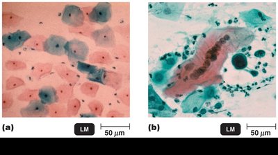
Example: The image above shows (a) normal cervical cells and (b) cancerous cervical cells, illustrating transformation by viral infection (e.g., HPV).
Baltimore Classification of Viral Genomes
The Baltimore classification groups viruses based on their genome type and replication strategy. Each class uses specific enzymes for genome replication and mRNA synthesis.
Class | Genome Type | Genome Replicated By | mRNA Generated By |
|---|---|---|---|
I | dsDNA | DNA polymerase | RNA polymerase |
II | +ssDNA | -ssDNA (by DNApol), then RNA polymerase | RNA polymerase |
III | dsRNA | RNA-dependent RNA polymerase (RdRp) | +ssRNA strand used as mRNA |
IV | +ssRNA | RNA-dependent RNA polymerase | Genome used directly as mRNA |
V | -ssRNA | +ssRNA (by RdRp), then used as mRNA | RNA-dependent RNA polymerase |
VI | +ssRNA (retrovirus) | Reverse transcriptase to DNA, then RNA polymerase | DNA (by RT), then RNA polymerase |
VII | dsDNA (with RNA intermediate) | RNA polymerase to RNA, then RT to dsDNA | RNA polymerase |
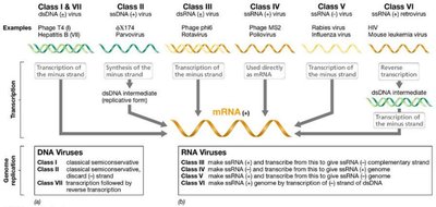
Example: The image above summarizes the Baltimore classification, showing genome types, replication, and transcription strategies for each class.
Key Enzymes: DNA polymerase, RNA polymerase, RNA-dependent RNA polymerase, and reverse transcriptase are essential for viral genome replication and mRNA synthesis.
Latent and Persistent Viral Infections
Latent Infection: Virus remains dormant within the host cell, with no active replication (e.g., herpes simplex virus).
Persistent Infection: Virus continuously replicates at low levels without causing cell death (e.g., hepatitis B virus).
Prions – Proteinaceous Infectious Particles
Genetic Material: Prions lack nucleic acids; they are composed solely of protein.
Structure: Abnormally folded proteins that induce misfolding of normal host proteins.
Diseases: Cause spongiform encephalopathies (e.g., Creutzfeldt-Jakob disease, mad cow disease).
Replication: Prion proteins convert normal proteins into the prion form.
Pathogenesis: Accumulation of prion proteins forms plaques in the CNS, leading to neuronal death and loss of function.
