 Back
BackViruses and Prions: Structure, Replication, and Clinical Relevance
Study Guide - Smart Notes
 Tailored notes based on your materials, expanded with key definitions, examples, and context.
Viruses and Prions
Introduction to Viruses
Viruses are submicroscopic, acellular infectious agents that require a host cell for replication. The study of viruses is known as virology. Viruses can infect all forms of life, including bacteria (bacteriophages), animals, and plants. They are considered nonliving because they lack cellular structure and metabolism.
Size: Typically 20–400 nm, much smaller than prokaryotic and eukaryotic cells.
Obligate intracellular pathogens: Cannot reproduce outside a host cell.
Host range: Can infect every branch in the tree of life.
Comparison of Viruses, Prokaryotes, and Eukaryotes
The following table summarizes the main differences between viruses, prokaryotes, and eukaryotes:
Characteristic | Viruses | Prokaryotes | Eukaryotes |
|---|---|---|---|
Cells? | No | Yes | Yes |
Considered alive? | No | Yes | Yes |
Relative size | Smaller than prokaryotes | Bigger than viruses, smaller than eukaryotes | Bigger than prokaryotes and viruses |
Filterable | Yes | Rarely | No |
Structure | Capsid and nucleic acid | Cells without nuclei | Cells with nuclei |
Replication | Hijack host machinery | Binary fission | Mitosis/Meiosis |
Metabolism | No | Yes | Yes |
Genome | DNA or RNA | DNA | DNA |
Virion Structure
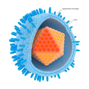
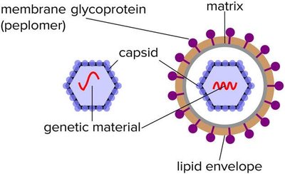
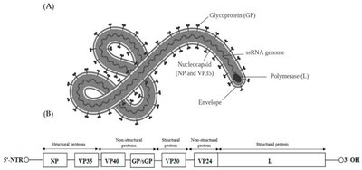
A virion is a single, infectious virus particle. It consists of a protective protein shell called a capsid, which encloses the viral genome (either DNA or RNA). Some viruses also possess an outer lipid envelope derived from the host cell membrane.
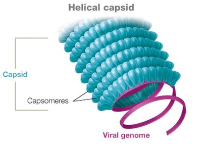
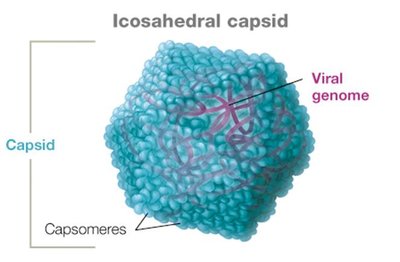
Capsid: Made of protein subunits called capsomeres.
Envelope: Lipid-based, present in some animal viruses.
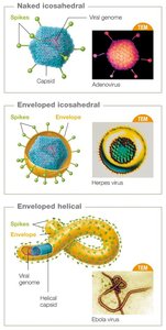
Capsid Symmetry
Animal viruses typically have either helical or icosahedral capsids, while some have complex structures.
Helical capsid: Hollow tube-like structure.
Icosahedral capsid: Three-dimensional polygonal structure.
Complex capsid: Found in some viruses like smallpox and bacteriophages.
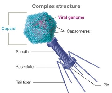
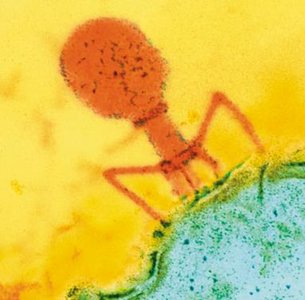
Bacteriophage Structure
Bacteriophages have complex capsids, often with icosahedral heads and additional structures for genome injection into bacteria.
Viral Envelopes and Spikes
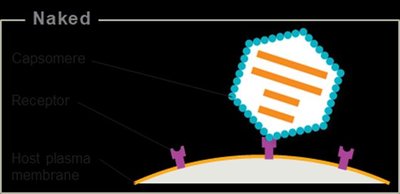
Some viruses are enveloped, possessing a lipid membrane with embedded glycoprotein spikes (peplomers) that facilitate host cell attachment and entry. Naked viruses lack this envelope.
Enveloped viruses: Acquire envelope by budding from host cell.
Naked viruses: Released by host cell lysis.
Spikes: Glycoproteins that determine host specificity and facilitate entry.
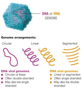
Viral Genomes
Viral genomes are highly variable and can be composed of DNA or RNA, which may be single- or double-stranded, linear, circular, or segmented. Most viruses have fewer than 300 genes, encoding structural proteins, enzymes, and replication factors.
Genome arrangements: Circular, linear, or segmented.
Types: ssDNA, dsDNA, ssRNA, dsRNA.
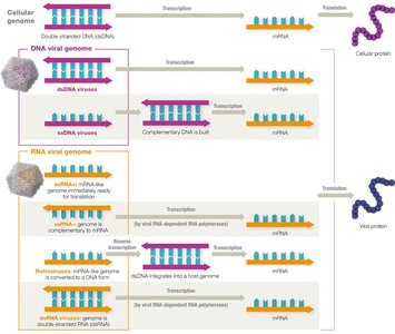
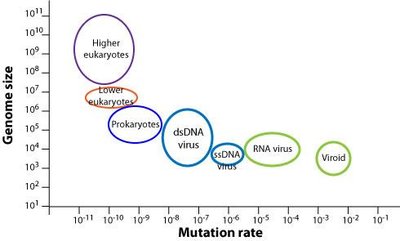
Viral Replication and Mutation
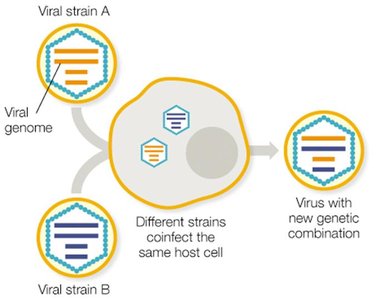
Viruses rely on host cell machinery for replication. Their genomes mutate rapidly, especially RNA viruses, due to lack of proofreading by RNA polymerases. This leads to high genetic variability and the emergence of new strains.
Mutation effects: Neutral, beneficial, or detrimental.
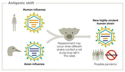
Reassortment: Exchange of genome segments between coinfecting viruses, leading to new strains.
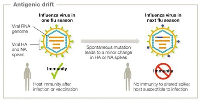
Antigenic Drift and Shift
Influenza viruses frequently undergo genetic changes in their surface antigens (HA and NA spikes), leading to antigenic drift (minor changes) and antigenic shift (major reassortment events). These processes can reduce immune recognition and lead to epidemics or pandemics.
Antigenic drift: Minor mutations in spike proteins.
Antigenic shift: Major reassortment, often resulting in new, highly infectious strains.
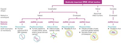
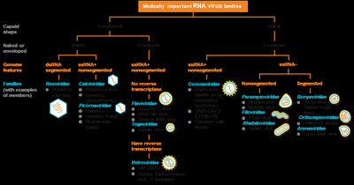
Classification and Naming of Viruses
Viruses are classified based on nucleic acid type, capsid symmetry, presence of envelope, and genome architecture. The highest taxonomic rank is phylum, followed by order, family, subfamily, genus, and species.
Taxon | Example | Naming Convention |
|---|---|---|
Order | Herpesvirales | Italicized, ends in 'virales' |
Family | Herpesviridae | Italicized, ends in 'viridae' |
Subfamily | Alphaherpesvirinae | Italicized, ends in 'virinae' |
Genus | Simplexvirus | Italicized, ends in 'virus' |
Species | Human herpesvirus-1 | Italicized, proper nouns capitalized |
Medically Important Virus Families
DNA and RNA viruses are grouped by genome type, capsid shape, and envelope presence. Examples include Herpesviridae, Papillomaviridae, Retroviridae, and Flaviviridae.
Host Range and Tropism
Host range is the spectrum of species a virus can infect, while tropism refers to the specific tissues or cells targeted. Some viruses have broad host ranges (e.g., Ebola), while others are highly specific (e.g., measles virus infects only humans).
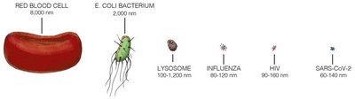
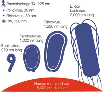
Virus Sizes
Viruses vary greatly in size, from small rhinoviruses (30 nm) to large pithoviruses (1,500 nm). Some bacteria are similar in size to large viruses.
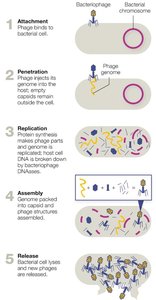
Bacteriophage Replication
Bacteriophages replicate via the lytic or lysogenic cycle. The lytic cycle results in host cell lysis and release of new phages, while the lysogenic cycle involves integration of the phage genome into the host chromosome (prophage).
Lytic cycle steps: Attachment, penetration, replication, assembly, release.
Lysogenic cycle: Phage genome integrates and can confer new properties (phage conversion).

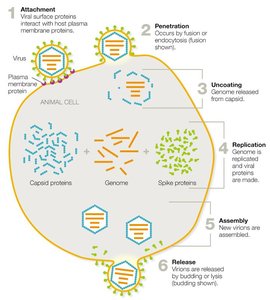
Animal Virus Replication
Animal viruses follow a general replication cycle: attachment, penetration, uncoating, replication, assembly, and release. Enveloped viruses are released by budding, while naked viruses cause cell lysis.
Persistent Infections and Oncogenic Viruses
Some animal viruses cause persistent infections, which may be chronic (continuous virion release, e.g., HIV) or latent (periods of dormancy, e.g., herpesviruses). Oncogenic viruses can induce cancer by disrupting cell cycle regulation.
Chronic infection: Continuous low-level replication (e.g., HIV).
Latent infection: Intermittent flare-ups (e.g., HSV-1, HSV-2, Varicella-zoster).
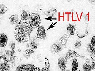
Oncogenic viruses: HPV, HTLV, Epstein–Barr virus, Hepatitis B and C viruses.
Virus | Genome | Integrates? | Cancer Link | Mechanism |
|---|---|---|---|---|
HPV | DNA | Yes | Cervical, oropharyngeal, anal, etc. | Uncontrolled cell division |
HTLV | RNA | Yes | Adult T-cell leukemia | Uncontrolled cell division |
Hepatitis B/C | DNA/RNA | No | Liver cancer | Chronic inflammation, DNA damage |
Virus Cultivation and Detection
Viruses are cultivated using bacterial cultures (for phages), embryonated eggs, tissue culture, or live animals. Detection methods include plaque assays, molecular techniques (PCR, sequencing), and immunological assays (ELISA, agglutination).
Plaque assay: Measures viral titer as plaque-forming units (PFUs).
ELISA: Detects viral antigens or antibodies via color change.
Agglutination: Latex beads clump in presence of viral antigen or antibody.
Nucleic acid detection: PCR, sequencing, fluorescent probes.
Antiviral Drugs and Vaccines
Antiviral drugs target various stages of the viral life cycle, including attachment, penetration, replication, and release. Most antivirals limit infection rather than cure it. Vaccines are crucial for prevention.
Entry inhibitors: Block viral attachment or fusion (e.g., docosanol, palivizumab).
Nucleoside analogs: Mimic nucleotides, inhibit replication (e.g., acyclovir, ribavirin).
Reverse transcriptase inhibitors: Target retroviruses (e.g., AZT).
Interferons: Boost host antiviral response.
Release inhibitors: Block virion budding (e.g., oseltamivir).
Prions
Prions are infectious proteins that lack nucleic acids. They cause transmissible spongiform encephalopathies (TSEs), such as Creutzfeldt-Jakob disease, by inducing misfolding of normal prion proteins in the brain, leading to neurodegeneration.
Transmission: Ingestion, inherited mutations, or sporadic misfolding.
Diagnosis: Detection of spongiform changes in brain tissue post-mortem.
Prion-like diseases: Some neurodegenerative diseases (e.g., Alzheimer's, Parkinson's) exhibit prion-like mechanisms.
Clinical Application: Case Study Questions
Screening old blood samples: Use molecular techniques (PCR, sequencing) and immunological assays (ELISA, agglutination) to detect new viruses.
Classifying a new virus: Determine nucleic acid type, capsid structure, envelope presence, and genome architecture using sequencing and electron microscopy.
Isolating and growing virus: Inoculate susceptible cell cultures or animal models, observe cytopathic effects, and confirm with molecular assays.
Antivirals for RNA viruses: Nucleoside analogs (e.g., ribavirin), reverse transcriptase inhibitors (for retroviruses), and entry inhibitors. These drugs inhibit replication or entry.
Shared genes and immune response: Strong immune reaction in vaccinated animals suggests antigenic similarity between the new virus and hepatitis C virus.
Low viral titers: Suggests a persistent, possibly latent or chronic infection with low-level replication.
Oncogenic potential: The absence of cancer in most infected individuals does not rule out oncogenicity; other factors (host genetics, co-infections) may influence cancer development.
