 Back
Viruses and Their Replication: Structure, Diversity, and Life Cycle
Study Guide - Smart Notes
 Tailored notes based on your materials, expanded with key definitions, examples, and context.
Viruses: Definition and Characteristics
What is a Virus?
Viruses are genetic elements that can replicate only inside a living cell, known as the host cell. Although viruses possess their own genome encoding functions necessary for replication, they are not considered living organisms because they lack independent metabolic activity and rely entirely on the host cell for energy, metabolic intermediates, and protein synthesis. Viral replication is only possible after successful entry into a suitable host cell, a process known as infection.
Viral Genomes: Diversity and Classification
Types of Viral Genomes
Viral genomes are highly diverse and can be composed of DNA or RNA, which may be single-stranded (ss) or double-stranded (ds). Single-stranded RNA viruses can be either positive-sense (+) or negative-sense (–). Genomes may be linear or circular, and some unusual viruses use both DNA and RNA at different stages of their life cycle. Viral genomes are typically much smaller than those of cellular organisms, with the smallest viruses containing fewer than 2,000 base pairs and only two genes.

Host Range and Types of Infection
Viruses are classified based on the hosts they infect, including bacteria (bacteriophages), archaea, protozoa, fungi (mycoviruses), animals, and plants. There are two main types of viral infection:
Lytic infection: Virus replicates and destroys the host cell, redirecting host metabolism to support viral replication and assembly. The host cell is lysed to release new virions.
Lysogenic infection: Viral genome becomes part of the host genome, genetically altering the host without destroying it.
Virus Structure and Virion Composition
Virion: The Extracellular Form
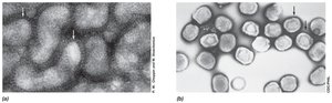
The virion is the extracellular form of the virus, enabling transmission between host cells. Virions are typically smaller than prokaryotic cells, ranging from 0.02 to 0.3 µm. The capsid is a protein shell that contains the viral genome, and together with the nucleic acid forms the nucleocapsid. Capsid proteins, called capsomeres, are arranged in a highly repetitive pattern around the nucleic acid.
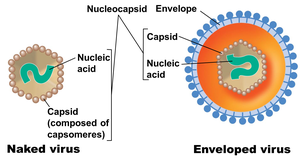
Most bacterial viruses are naked viruses (no further layers), while many animal viruses have an envelope (outer layer of protein and lipid).
Symmetry in Virion Structure
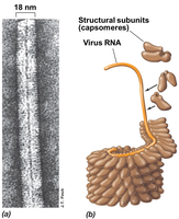
Helical symmetry: Rod-shaped viruses, such as Tobacco mosaic virus (TMV), have capsids arranged in a helix. The length is determined by the nucleic acid, and the width by the size and packaging of protein subunits.
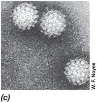
Icosahedral symmetry: Spherical viruses have capsids arranged in 20 triangular faces, with clusters of capsomeres forming each face. Most have 240–360 capsomeres.
Complex Virion Structures
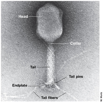
Some viruses, such as bacteriophage T4, have complex structures with an icosahedral head and a helical tail. The tail is composed of multiple proteins and is essential for attachment and DNA injection into the host cell.
Enveloped Viruses
Enveloped viruses have a membrane surrounding the nucleocapsid, typically derived from the host cell membrane as the virus exits the cell. The envelope assists in infection by fusing with the host membrane, allowing the entire virion to enter the cell.
Viral Life Cycle
Steps of the Viral Life Cycle
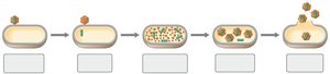
The viral life cycle in a permissive host cell consists of five main steps:
Attachment (adsorption): Virus binds to specific receptors on the host cell.
Entry: Virion or its nucleic acid enters the host cell.
Synthesis: Viral nucleic acid and proteins are synthesized using host cell machinery.
Assembly: Capsids are assembled and viral genomes are packaged into new virions.
Release: Mature virions are released from the host cell, often by lysis.
Attachment Specificity
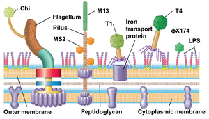
Attachment is highly specific, requiring viral proteins to interact with host cell receptors. If the receptor is absent or altered, infection cannot occur. Receptors may be proteins, carbohydrates, glycoproteins, lipids, or cell structures, and often serve normal functions for the host cell.
Bacteriophage T4 Infection Mechanism
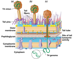
Bacteriophage T4 infects Escherichia coli by attaching via tail fibers to LPS on the cell surface. Tail fibers retract, tail pins contact the cell wall, and a lysozyme-like enzyme forms a pore in the peptidoglycan. The tail sheath contracts, injecting viral DNA into the cytoplasm.
Transcription, Translation, and Viral Assembly
Phage T4 Gene Expression
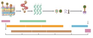
After T4 entry, host DNA and RNA synthesis ceases, and phage gene transcription begins. The T4 genome encodes three sets of proteins:
Early proteins: Enzymes for DNA replication and modification of host RNA polymerase.
Middle proteins: Additional proteins for RNA polymerase modification and expression of late genes.
Late proteins: Structural proteins for head and tail, and enzymes for virion release.
Virus Growth Curve
One-Step Growth Curve
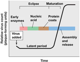
Viral replication follows a "one-step" growth curve, with no increase in virion numbers until cell lysis. The latent period includes the eclipse (no detectable virions) and maturation (packaging of nucleic acids into capsids). Burst size refers to the number of virions released per cell.
Culturing, Detecting, and Counting Viruses
Plaque Assay
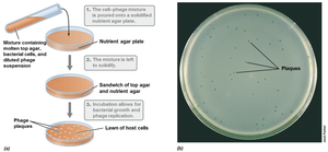
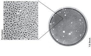
The plaque assay is used to determine viral titer (number of infectious virions per volume). Plaques are clear zones on lawns of host cells, each resulting from infection by a single virus particle. Viral titer is reported as "plaque-forming units" per mL.
Plating Efficiency
The number of plaque-forming units is usually lower than direct counts by electron microscopy due to inactive virions, suboptimal conditions, or damage. Bacterial viruses typically have efficiencies >50%, while animal viruses may be much lower (0.1–1%).
Temperate Bacteriophages and Lysogeny
Lytic vs. Lysogenic Pathways
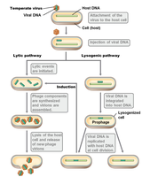
Temperate viruses can alternate between:
Lytic pathway: Virion assembly and host cell lysis.
Lysogenic pathway: Viral genome integrates into host chromosome and replicates with it, conferring new genetic properties (lysogenic conversion).
Bacteriophage Lambda
Bacteriophage lambda infects E. coli with a linear, double-stranded DNA genome. Upon entry, the DNA circularizes and may integrate into the host chromosome at the att site, requiring lambda integrase. The decision between lysis and lysogeny depends on the levels of two repressor proteins, cI and Cro. Accumulation of cI leads to lysogeny, while Cro accumulation leads to lysis.
Bacterial Virus Diversity
Genome Types and Replication
Most bacteriophages are head-and-tail with double-stranded DNA genomes. Single-stranded DNA viruses convert their genome into a double-stranded form for transcription and replication. Some, like MS2, have a plus-sense ssRNA genome that can be translated directly and encode RNA replicase for genome replication. Small viral genomes may contain overlapping genes.
Animal Viruses: Overview
Key Differences from Bacterial Viruses
The entire virion enters the animal cell.
Many animal viruses replicate in the nucleus.
All types of viral genomes exist, classified as enveloped or nonenveloped.
Viral Ecology and the Virosphere
Environmental Abundance and Impact
Viruses are ubiquitous in all environments, often outnumbering prokaryotes. Bacteriophages significantly impact bacterial evolution through lysogenic conversion and transduction. Most of Earth's genetic diversity resides in viruses, and many viral genes have unknown functions.
Additional info: Academic context was added to clarify definitions, mechanisms, and examples for completeness and exam preparation.
