 Back
BackViruses: Structure, Classification, Multiplication, and Pathogenicity
Study Guide - Smart Notes
 Tailored notes based on your materials, expanded with key definitions, examples, and context.
General Characteristics of Viruses
Obligatory Intracellular Parasites
Viruses are unique infectious agents that require living host cells to multiply. They lack the cellular machinery necessary for independent life and reproduction.
Contain either DNA or RNA: Viruses possess a single type of nucleic acid, either DNA or RNA, but never both.
No ribosomes: Viruses cannot synthesize proteins independently.
No ATP-generating mechanism: Viruses rely on host cells for energy.
Contain a protein coat: The protein coat, or capsid, protects the viral nucleic acid.
Some viruses are enclosed by an envelope: This lipid membrane is derived from the host cell.
Some viruses have spikes: Spikes are glycoproteins that aid in attachment to host cells.
Host range: Most viruses infect only specific types of cells in one host, determined by attachment sites and cellular factors.
Virion Structure
Components of a Virion
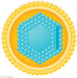
A virion is a complete, infectious virus particle. Its structure is essential for protection and delivery of the viral genome.
Nucleic acid: DNA or RNA, single- or double-stranded.
Capsid: Protein shell composed of subunits called capsomeres.
Envelope: Lipid membrane surrounding some viruses.
Spikes: Surface projections for attachment.
Virus Morphology
Viruses are classified by their shape and structure:
Polyhedral: Many-sided, often icosahedral.
Helical: Rod-shaped, with nucleic acid inside a helical capsid.
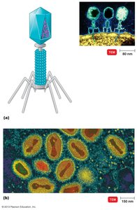
Complex: More elaborate structures, such as bacteriophages.
Polyhedral Viruses
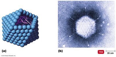
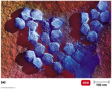
Polyhedral viruses, such as Mastadenovirus, have a capsid with many sides, typically icosahedral.
Example: Mastadenovirus
Enveloped Helical Viruses
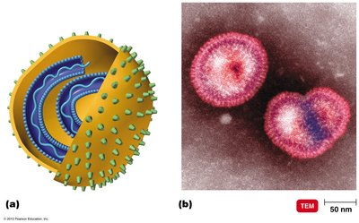
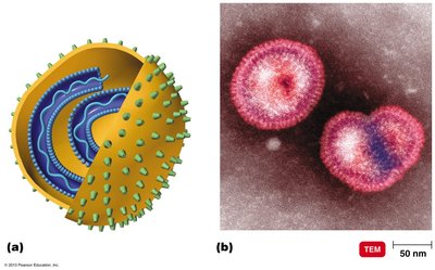
Enveloped helical viruses, such as Influenzavirus, have a helical capsid surrounded by an envelope.
Example: Influenzavirus
Helical Viruses
Helical viruses, such as Ebola virus, have a cylindrical capsid.
Example: Ebola virus
Complex Viruses
Complex viruses, such as T-even bacteriophages and Orthopoxvirus, have intricate structures with additional components like tails and fibers.
Example: T-even bacteriophage, Orthopoxvirus
Taxonomy of Viruses
Classification Principles
Virus taxonomy is based on genetic material, host range, and structural features.
Family names: End in -viridae
Genus names: End in -virus
Viral species: Group sharing genetic information and ecological niche
Common names: Used for species
Subspecies: Designated by numbers
Examples of Virus Families
Herpesviridae: Includes Herpesvirus (HHV-1, HHV-2, HHV-3)
Retroviridae: Includes Lentivirus (HIV-1, HIV-2)
Isolation, Cultivation, and Identification of Viruses
Growing Viruses
Viruses must be grown in living cells, as they cannot replicate independently.
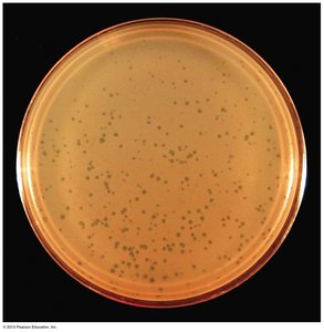
Bacteriophages: Form plaques on a lawn of bacteria.
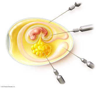
Animal viruses: Grown in living animals, embryonated eggs, or cell cultures.
Continuous cell lines: Transformed cells that can be maintained indefinitely.
Viral Multiplication
The Lytic Cycle
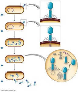
The lytic cycle is a process by which bacteriophages replicate and destroy the host cell.
Attachment: Phage attaches to host cell via tail fibers.
Penetration: Phage lysozyme opens cell wall; DNA injected.
Biosynthesis: Production of phage DNA and proteins.
Maturation: Assembly of phage particles.
Release: Cell wall breaks, releasing new virions.
The Lysogenic Cycle
In the lysogenic cycle, phage DNA integrates into the host genome and replicates without killing the cell.
Prophage DNA: Incorporated in host DNA.
Phage conversion: Host cell acquires new properties.
Specialized transduction: Transfer of specific bacterial genes by phage.
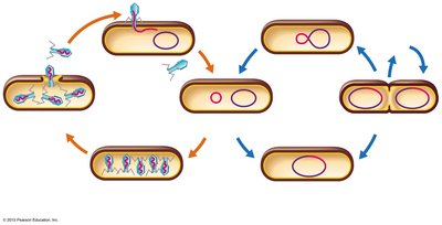
Transduction by a Bacteriophage
Transduction is the process by which bacterial DNA is transferred from one cell to another by a virus.
Generalized transduction: Any bacterial gene may be transferred.
Specialized transduction: Only specific genes are transferred.
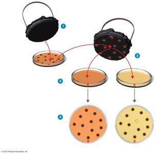
Multiplication of Animal Viruses
Steps in Animal Virus Multiplication
Animal viruses follow a series of steps to infect host cells:
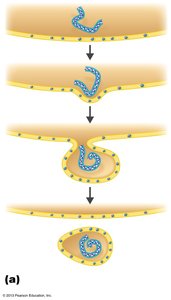
Attachment: Viruses attach to cell membrane.
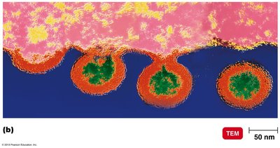
Penetration: Entry by endocytosis or fusion.
Uncoating: Viral or host enzymes remove capsid.
Biosynthesis: Production of nucleic acid and proteins.
Maturation: Assembly of nucleic acid and capsid proteins.
Release: By budding (enveloped viruses) or rupture.
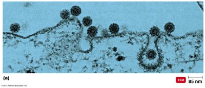
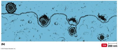
Replication of DNA-Containing Animal Viruses
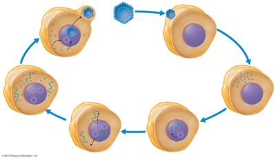
DNA viruses replicate in the host cell nucleus and follow a specific sequence of events.
Attachment, Entry, and Uncoating
Biosynthesis: Early and late transcription/translation
Maturation and Release
Replication of RNA-Containing Animal Viruses
RNA viruses replicate in the cytoplasm and use viral RNA-dependent RNA polymerase.
+ strand (sense): Acts as mRNA
- strand (antisense): Must be transcribed to + strand
dsRNA: Double-stranded RNA viruses
Multiplication and Inheritance of Retroviridae
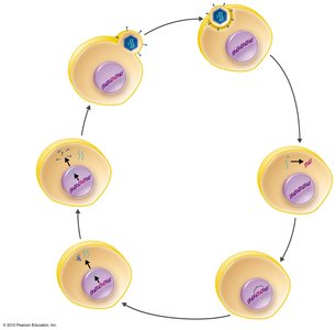
Retroviruses use reverse transcriptase to produce DNA from RNA, which integrates into the host genome.
Reverse transcriptase: Enzyme that synthesizes DNA from RNA
Integration: Viral DNA becomes a provirus in host chromosome
Transcription: Provirus DNA transcribed to produce new viral genomes and proteins
Release: Virus buds from host cell, acquiring envelope
Latent and Persistent Viral Infections
Latent and Persistent/Chronic Infections
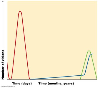
Some viruses remain dormant in host cells, while others cause prolonged disease.
Latent infection: Virus remains in asymptomatic host cell for long periods (e.g., cold sores, shingles)
Persistent infection: Disease process occurs over a long period, often fatal (e.g., subacute sclerosing panencephalitis)
Viruses and Cancer
Oncogenic Viruses
Oncogenic viruses can cause cancer by integrating their genetic material into host DNA and transforming normal cells.
Activated oncogenes: Transform normal cells into cancerous cells
Transformed cells: Increased growth, loss of contact inhibition, tumor-specific antigens
Integration: Genetic material of oncogenic viruses becomes part of host DNA
Oncogenic DNA viruses: Adenoviridae, Herpesviridae, Poxviridae, Papovaviridae, Hepadnaviridae
Oncogenic RNA viruses: Retroviridae (HTLV-1, HTLV-2)
Virus Families That Affect Humans
Major Virus Families
Adenoviridae: Double-stranded DNA, nonenveloped; respiratory infections, tumors
Papovaviridae: Double-stranded DNA, nonenveloped; papillomavirus, some serotypes cause cancer

Poxviridae: Double-stranded DNA, enveloped; smallpox, cowpox
Herpesviridae: Double-stranded DNA, enveloped; includes HHV-1 to HHV-8, some remain latent
Hepadnaviridae: Double-stranded DNA, enveloped; hepatitis B virus, uses reverse transcriptase
Picornaviridae: Single-stranded RNA, + strand, nonenveloped; enterovirus, rhinovirus, hepatitis A
Caliciviridae: Single-stranded RNA, + strand, nonenveloped; hepatitis E, norovirus
Flaviviridae: Single-stranded RNA, + strand, enveloped; arboviruses, hepatitis C
Rhabdoviridae: Single-stranded RNA, − strand; rabies virus
Filoviridae: Single-stranded RNA, − strand; Ebola, Marburg viruses
Orthomyxoviridae: Single-stranded RNA, − strand, multiple strands; influenza viruses
Retroviridae: Single-stranded RNA, two strands; HIV, oncogenic viruses
Antigenic Shift and Independent Assortment
Antigenic Shift in Influenza Virus
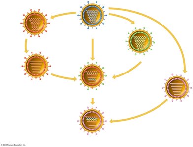
Antigenic shift is the process by which two different strains of a virus combine to form a new subtype with mixed surface antigens.
Mechanism: Independent assortment of viral genome segments
Example: 2009 H1N1 pandemic resulted from reassortment of human, avian, and swine influenza viruses
Prions
Proteinaceous Infectious Particles
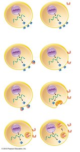
Prions are infectious proteins that cause neurodegenerative diseases.
Inherited and transmissible: By ingestion, transplant, and surgical instruments
Spongiform encephalopathies: Includes sheep scrapie, Creutzfeldt-Jakob disease, mad cow disease
Cellular Prion Protein (PrPC): Normal protein on cell surface
Scrapie Protein (PrPSc): Abnormal form, accumulates in brain cells, forming plaques
Smallpox
Smallpox Virus and Disease
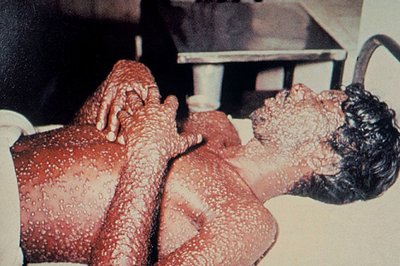
Smallpox, caused by Variola virus, was a deadly disease eradicated by vaccination.
Symptoms: Maculopapular rash, raised fluid-filled blisters
Eradication: Last natural outbreak in the US in 1949; declared eliminated in 1980
 ----------------------------------------
