 Back
BackViruses: Structure, Morphology, and Cultivation
Study Guide - Smart Notes
 Tailored notes based on your materials, expanded with key definitions, examples, and context.
Viruses: Structure, Morphology, and Cultivation
Introduction to Viruses
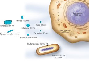
Viruses, also known as virions or virus particles, are acellular infectious agents that require a host cell for replication. They are significantly smaller than their host cells and exhibit a wide range of structural diversity.
Size: Viruses typically range from 20 to 400 nm in diameter, making them much smaller than most prokaryotic and eukaryotic cells.
Host Range: Viruses can infect all forms of life, including bacteria, archaea, plants, animals, and fungi.
Viral Structure
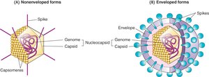
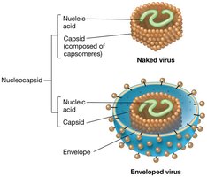
Nucleocapsid and Capsid
The fundamental structure of a virus is the nucleocapsid, which consists of the viral genome (nucleic acid) surrounded by a protective protein coat called the capsid. The capsid is composed of protein subunits known as capsomeres.
Capsomeres: Protein subunits that self-assemble to form the capsid, providing protection and symmetry to the virion.
Symmetry: Capsids can exhibit helical, icosahedral, or complex symmetry.
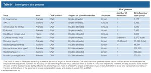
Viral Genome
Viral genomes are highly diverse and can be composed of either DNA or RNA, which may be single-stranded or double-stranded, linear or circular, and segmented or non-segmented. The genome encodes the information necessary for viral replication and assembly.
Virus | Host | DNA or RNA | Single- or Double-stranded | Structure | Number of molecules | Size (bases or base pairs) |
|---|---|---|---|---|---|---|
HPV (human papillomavirus) | Animals | DNA | Double-stranded | Circular | 1 | 7,900 |
Influenza virus | Animals | RNA | Single-stranded | Linear | 8 | 13,588 |
Bacteriophage T4 | Bacteria | DNA | Double-stranded | Linear | 1 | 168,903 |
Human immunodeficiency virus (HIV) | Animals | RNA | Single-stranded | Linear | 2 | 9,749 |
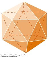
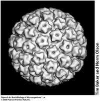
Capsid Symmetry
Capsids can be arranged in different symmetrical forms, which influence the overall shape and size of the virus:
Helical Symmetry: Protein subunits assemble in a spiral around the nucleic acid, forming rod-shaped virions. The length of the virus is determined by the length of the nucleic acid.
Icosahedral Symmetry: The capsid forms a roughly spherical structure composed of 20 equilateral triangular faces, providing maximum internal volume for the genome.
Viral Structure Extras: Envelopes and Complex Virions
Some viruses possess additional structural features:
Envelope: A lipid bilayer derived from the host cell membrane that surrounds the capsid in some viruses. Enveloped viruses often have surface proteins (spikes) involved in host recognition and entry.
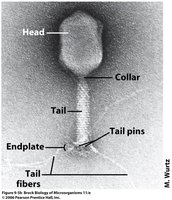
Complex Virions: Some viruses, such as bacteriophages, have complex structures with additional components like tails, tail fibers, and base plates for host attachment and genome delivery.
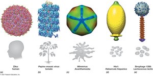
Virion Morphology and Their Hosts
Viruses display a variety of morphologies, which are often related to their host range and mode of infection. Common morphologies include helical, icosahedral, complex, and enveloped forms.
Enzymes Inside the Nucleocapsid
Some viruses package essential enzymes within their nucleocapsid to facilitate replication and infection:
Bacteriophage T4 lysozyme: Degrades bacterial cell walls during infection.
RNA replicases: Required by RNA viruses for genome replication.
Reverse transcriptase, protease, integrase: Found in retroviruses, these enzymes are necessary for reverse transcription, protein processing, and integration into the host genome.
Extracellular vs. Intracellular Forms
Viruses exist in two distinct forms:
Extracellular (Virion): The inert, infectious particle outside the host cell, incapable of metabolic activity.
Intracellular: The active form inside the host cell, where the virus hijacks the host's metabolic machinery to replicate and assemble new virions.
Culturing, Detecting, and Counting Viruses
Methods for Culturing Viruses
Viruses require living cells for propagation. The methods for culturing viruses depend on the host type:
Bacterial Viruses (Bacteriophages): Grown using bacterial cultures in liquid medium or as lawns on agar plates.
Animal and Plant Viruses: Cultivated in tissue cultures derived from animal organs or plant tissues. Plant viruses may require specialized systems such as hairy root cultures.
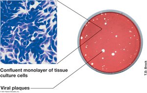
Detecting and Counting Viruses: The Plaque Assay
The plaque assay is a standard method for quantifying infectious virus particles. It involves infecting a lawn of host cells with diluted virus and counting the resulting clear zones (plaques) where cells have been lysed by viral infection.
Titer: The concentration of infectious virions, expressed as plaque-forming units (PFU) per volume.
Calculation: Titer is determined by counting plaques and accounting for dilution factors, analogous to counting bacterial colonies.
Animal Viruses: Similar plaque assays are used, but with tissue culture cells.
Plant Viruses: More challenging to quantify; often require purification and microscopic or protein-specific detection methods.

Summary Table: Key Features of Viruses
Feature | Description |
|---|---|
Size | 20–400 nm (smaller than host cells) |
Genome | DNA or RNA; single- or double-stranded; linear or circular |
Capsid | Protein shell made of capsomeres; helical or icosahedral symmetry |
Envelope | Lipid bilayer derived from host (in some viruses) |
Replication | Obligate intracellular parasites; require host cell machinery |
Detection | Plaque assay, microscopy, protein-specific methods |
