 Back
BackViruses, Viroids, and Prions: Structure, Classification, and Replication
Study Guide - Smart Notes
 Tailored notes based on your materials, expanded with key definitions, examples, and context.
Characteristics of Viruses
Definition and General Properties
Viruses are minuscule, acellular infectious agents that contain either DNA or RNA as their genetic material. They are responsible for a wide range of diseases in humans, animals, plants, and even bacteria. Unlike cellular life forms, viruses cannot carry out metabolic processes, do not grow or respond to their environment, and cannot reproduce independently. Instead, they rely on host cells to replicate and propagate.
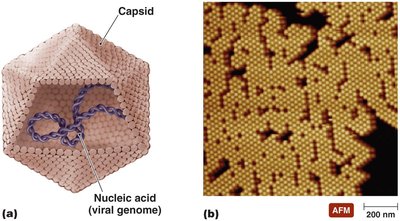
Virion: The extracellular, infectious state of a virus, consisting of a nucleic acid genome surrounded by a protein coat (capsid).
Intracellular State: Once inside a host cell, the capsid is removed, and the virus exists as nucleic acid.
Lack of Cellular Structures: Viruses do not possess cytoplasmic membranes, cytosol, or organelles (with rare exceptions).
Genetic Material of Viruses
Viruses exhibit remarkable diversity in their genetic material, which is a primary criterion for their classification. Their genomes may be composed of DNA or RNA, but never both, and can be single-stranded (ss) or double-stranded (ds), linear or circular, and segmented or non-segmented. Viral genomes are much smaller than those of cellular organisms.
Host Range and Specificity
Most viruses are highly specific, infecting only particular cell types within specific hosts due to the precise interaction between viral surface proteins and host cell receptors. Some viruses, termed generalists, can infect multiple cell types or species.
Size and Morphology
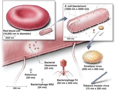
Viruses are among the smallest infectious agents, typically ranging from 20 to 300 nanometers. Their size is much smaller than most bacteria and eukaryotic cells.
Structure of Viruses
Capsid Morphology
The capsid is a protein shell that encases the viral genome, providing protection and facilitating attachment to host cells. Capsids are composed of subunits called capsomeres, which may be made of one or several types of proteins. The shape of the capsid can be helical, polyhedral (icosahedral), or complex.
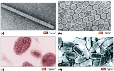
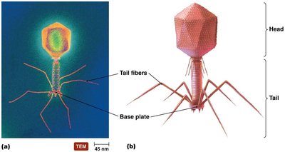
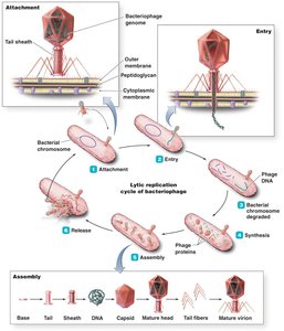
Complex Viruses: Bacteriophages
Bacteriophages are viruses that infect bacteria and often exhibit complex structures, including a head, tail, and tail fibers, which facilitate attachment and injection of genetic material into bacterial cells.
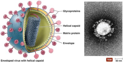
Enveloped Viruses
Some animal viruses possess an envelope derived from the host cell membrane, embedded with viral glycoproteins (spikes) that play a crucial role in host recognition and attachment. The envelope provides additional protection and aids in the infection process.
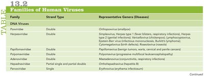
Classification of Viruses
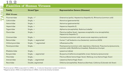
Families of Human Viruses
Viruses are classified based on their nucleic acid type (DNA or RNA), strandedness (single or double), and replication strategy. The following tables summarize the major families of DNA and RNA viruses and their representative diseases.
Family | Strand Type | Representative Genera (Diseases) |
|---|---|---|
Parvoviridae | Single | Erythrovirus (erythema infectiosum) |
Herpesviridae | Double | Simplexvirus (herpes), Varicellovirus (chickenpox), Epstein-Barr virus (mononucleosis, Burkitt's lymphoma) |
Poxviridae | Double | Orthopoxvirus (smallpox) |
Hepadnaviridae | Partial single and partial double | Orthohepadnavirus (hepatitis B) |
Family | Strand Type | Representative Genera (Diseases) |
|---|---|---|
Picornaviridae | Single, + | Enterovirus (poliovirus), Hepatovirus (hepatitis A), Rhinovirus (common cold) |
Flaviviridae | Single, + | Flavivirus (yellow fever), Hepacivirus (hepatitis C) |
Orthomyxoviridae | Single, - | Influenzavirus (influenza) |
Reoviridae | Double | Rotavirus (diarrhea) |
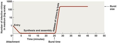
Viral Replication
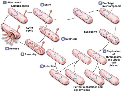
Lytic Replication Cycle
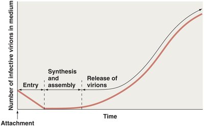
The lytic cycle is a replication process that results in the destruction (lysis) of the host cell. It consists of five main stages:
Attachment: Virus binds to specific receptors on the host cell surface.
Entry: Viral genome enters the host cell.
Synthesis: Host machinery is used to synthesize viral components.
Assembly: New virions are assembled from synthesized components.
Release: Host cell lyses, releasing new virions.
Lysogenic Replication Cycle
Some bacteriophages undergo lysogeny, a modified replication cycle where the viral genome integrates into the host chromosome as a prophage. The host cell continues to live and divide, passing the prophage to daughter cells. Environmental triggers can induce the prophage to enter the lytic cycle.
Replication of Animal Viruses
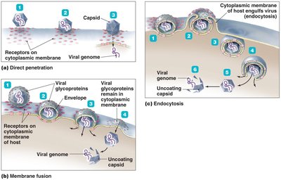
Animal viruses follow similar replication steps as bacteriophages but differ due to the presence of envelopes, the eukaryotic nature of host cells, and the absence of a cell wall. Entry mechanisms include direct penetration, membrane fusion, and endocytosis. The site of synthesis and assembly depends on the type of viral genome.
Attachment: Mediated by glycoprotein spikes or other molecules.
Entry: Can occur via direct penetration, membrane fusion, or endocytosis.
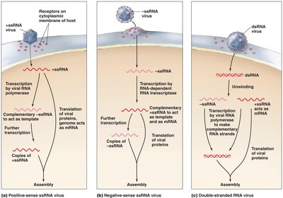
Synthesis: DNA viruses often replicate in the nucleus; RNA viruses in the cytoplasm.
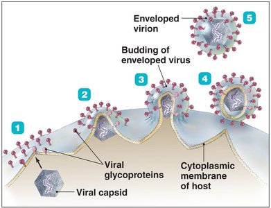
Assembly and Release: Enveloped viruses bud from the cell membrane; naked viruses are released by lysis or exocytosis.
Latency in Animal Viruses
Some animal viruses can remain dormant within host cells for extended periods (latency). Latent viruses may integrate into the host genome (provirus), and this incorporation is permanent. Latency can last for years without symptoms.
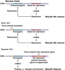
The Role of Viruses in Cancer
Oncogenes and Cancer Induction
Viruses can contribute to cancer development by carrying oncogenes, promoting host oncogenes, or interfering with tumor suppressor genes. Environmental factors such as UV light, radiation, and carcinogens also play a role. Examples of virus-induced cancers include Burkitt's lymphoma, Hodgkin's disease, Kaposi's sarcoma, and cervical cancer.
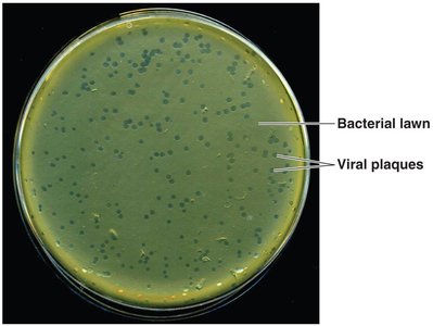
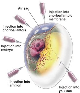
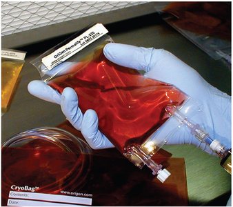
Culturing Viruses in the Laboratory
Methods of Culturing Viruses
In Mature Organisms: Viruses can be grown in bacteria, plants, or animals.
In Embryonated Chicken Eggs: Eggs provide a sterile, nutrient-rich environment for viral growth.
In Cell (Tissue) Culture: Cells isolated from organisms are grown in vitro. Two types are diploid cell cultures and continuous cell cultures.
Are Viruses Alive?
The debate over whether viruses are alive centers on their lack of independent metabolism and cellular structure. Some consider them complex pathogenic chemicals, while others view them as the simplest living entities due to their ability to invade cells, control host machinery, and replicate.
Other Parasitic Particles: Viroids and Prions
Viroids
Viroids are extremely small, circular RNA molecules that infect plants. They lack a protein capsid and may appear linear due to hydrogen bonding. Viroids are responsible for several plant diseases.
Prions
Prions are infectious proteins that cause fatal neurodegenerative diseases. The normal cellular form (PrPC) contains alpha-helices, while the disease-causing form (PrPSc) contains beta-sheets. Prion PrP induces conformational changes in normal PrP, leading to aggregation and brain damage. Prion diseases include BSE (mad cow disease), vCJD, and kuru. Prions are highly resistant to destruction and can only be inactivated by incineration or autoclaving in strong alkali.
