 Back
BackViruses, Viroids, and Prions: Structure, Classification, and Multiplication
Study Guide - Smart Notes
 Tailored notes based on your materials, expanded with key definitions, examples, and context.
General Characteristics of Viruses
Obligate Intracellular Parasites
Viruses are obligate intracellular parasites, meaning they require living host cells to multiply. Unlike most bacteria, viruses cannot reproduce or carry out metabolic processes outside a host cell. Some bacteria, such as Chlamydia and Rickettsia, also require host cells for replication, but they differ fundamentally from viruses in structure and metabolism.
Genetic Material: Viruses contain either DNA or RNA, but never both.
Protein Coat: All viruses have a protein coat (capsid) that protects their genetic material.
No Ribosomes: Viruses lack ribosomes and cannot synthesize proteins independently.
No ATP-Generating Mechanism: Viruses do not generate ATP and rely entirely on the host cell's machinery.
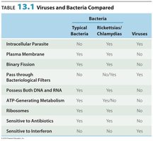
Table Purpose: This table compares the fundamental differences between typical bacteria, Rickettsias/Chlamydias, and viruses, highlighting the unique features of viruses such as the absence of ribosomes, ATP-generating metabolism, and their sensitivity to interferon.
Host Range and Viral Size
Host Specificity
The host range of a virus refers to the spectrum of host cells it can infect. Most viruses are highly specific, infecting only certain cell types within a single host species. This specificity is determined by the presence of compatible attachment sites on the host cell and appropriate cellular factors.
Examples: HIV infects CD4+ T cells; Hepatitis C virus targets hepatocytes via LDL receptors.
Bacteriophages: Viruses that infect bacteria are called bacteriophages or "phages."
Size: Viruses range from 20 nm to 1000 nm, much smaller than most bacteria.
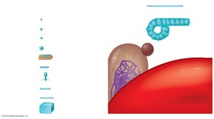
Image Purpose: This image illustrates the relative sizes of various viruses, a bacterium, and a human red blood cell, emphasizing the small size of viruses compared to cellular organisms.
Viral Structure
Basic Components
A virion is a complete, fully developed viral particle. The main structural components of a virion include:
Nucleic Acid: Can be DNA or RNA, single- or double-stranded, linear or circular.
Capsid: Protein coat composed of subunits called capsomeres.
Envelope: Some viruses possess a lipid, protein, and carbohydrate envelope derived from the host cell membrane.
Spikes: Glycoprotein projections from the envelope or capsid, important for attachment to host cells.
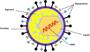
Image Purpose: This diagram shows the structural components of an enveloped virus, including the envelope, tegument, capsid, viral DNA/RNA, and glycoprotein spikes.
Types of Viral Morphology
Helical Viruses: Hollow, cylindrical capsid (e.g., tobacco mosaic virus).
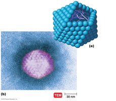
Polyhedral Viruses: Many-sided, typically icosahedral (e.g., adenovirus).
Enveloped Viruses: Surrounded by a lipid membrane (e.g., influenza virus).
Complex Viruses: Complicated structures, such as bacteriophages with additional components.
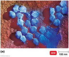
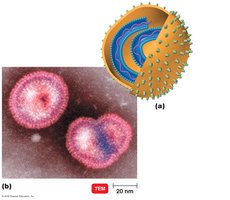
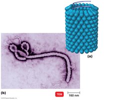
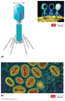
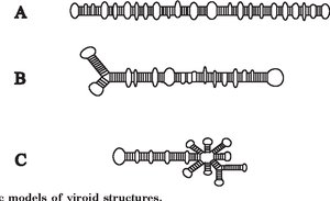
Viroids: Infectious agents composed solely of a short strand of circular, single-stranded RNA without a protein coat. They are the smallest known infectious pathogens.
Taxonomy of Viruses
Naming and Classification
Viruses are classified based on their genetic material, structure, and host range. The taxonomic hierarchy includes:
Order: Names end in -ales (e.g., Herpesvirales).
Family: Names end in -viridae (e.g., Herpesviridae).
Genus: Names end in -virus (e.g., Simplexvirus).
Species: Group of viruses sharing the same genetic information and ecological niche.
Subspecies: Designated by numbers (e.g., Herpes simplex virus 1, 2, 3).
Isolation, Cultivation, and Identification of Viruses
Growing Bacteriophages
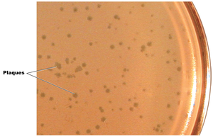
Bacteriophages are grown in bacterial cultures. They form plaques—clear zones on a bacterial lawn—each representing a single virus. The number of plaques can be quantified as plaque-forming units (PFU).
Growing Animal Viruses
In Living Animals: Used for studying pathogenesis and immune responses.
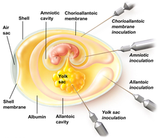
In Embryonated Eggs: Virus is injected into various egg compartments; growth is detected by embryo changes or death.
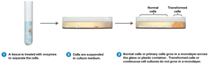
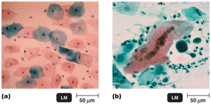
In Cell Cultures: Tissues are enzymatically dissociated, and cells are grown in culture. Viral infection is detected by cytopathic effects (CPE), such as cell rounding or detachment. Continuous cell lines are often used for routine virus culture.
Viral Identification
Cytopathic Effects: Observable changes in host cells due to viral infection.
Serological Tests: Detection of viral antigens or antibodies (e.g., Western blotting).
Nucleic Acid Techniques: PCR and RFLP analysis for viral genome identification.
Viral Multiplication
General Steps
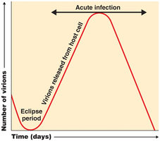
For a virus to multiply, it must invade a host cell and commandeer the host's metabolic machinery. The process is often depicted as a one-step growth curve, with an eclipse period followed by a burst of virion release.
Multiplication of Bacteriophages
Lytic Cycle: The phage replicates and lyses the host cell, releasing new virions.
Lysogenic Cycle: The phage DNA integrates into the host genome as a prophage and replicates passively with the host cell.
Lytic Cycle Steps (T-even Bacteriophages)
Attachment: Phage attaches to host cell.
Penetration: Phage injects DNA into host.
Biosynthesis: Synthesis of viral components.
Maturation: Assembly of new virions.
Release: Host cell lyses, releasing virions.
Lysogenic Cycle (Bacteriophage Lambda)
Phage DNA integrates into host DNA as a prophage.
Prophage is replicated with host genome.
Phage may later excise and enter the lytic cycle.
Phage conversion can result in new properties for the host cell.
Multiplication of Animal Viruses
Attachment: Virus binds to cell membrane receptors.
Entry: By receptor-mediated endocytosis or membrane fusion.
Uncoating: Viral genome is released inside the cell.
Biosynthesis: Synthesis of viral nucleic acids and proteins.
Maturation: Assembly of viral components.
Release: By budding (enveloped viruses) or cell rupture (nonenveloped viruses).
Biosynthesis of DNA and RNA Viruses
DNA Viruses
Replicate DNA in the host nucleus using viral enzymes.
Synthesize capsid proteins in the cytoplasm using host enzymes.
Major Families and Examples
Adenoviridae: dsDNA, nonenveloped; respiratory infections.
Poxviridae: dsDNA, enveloped; smallpox, vaccinia.
Herpesviridae: dsDNA, enveloped; herpes simplex, varicella-zoster, cytomegalovirus.
Papovaviridae: dsDNA, nonenveloped; papillomavirus (warts, HPV).
Hepadnaviridae: dsDNA, enveloped; hepatitis B virus (uses reverse transcriptase).
RNA Viruses
Replicate in the host cytoplasm using RNA-dependent RNA polymerase.
+ (Sense) Strand: Viral RNA serves directly as mRNA.
– (Antisense) Strand: Viral RNA must be transcribed to + strand before translation.
dsRNA: Both strands present; mRNA is transcribed from the – strand.
Major Families and Examples
Picornaviridae: ssRNA (+), nonenveloped; poliovirus, rhinovirus, hepatitis A.
Togaviridae: ssRNA (+), enveloped; rubella, chikungunya.
Rhabdoviridae: ssRNA (–), enveloped; rabies virus.
Reoviridae: dsRNA, nonenveloped; rotavirus.
Retroviruses
Single-stranded RNA viruses that produce DNA using reverse transcriptase.
Viral DNA integrates into the host genome as a provirus (e.g., HIV).
Viruses and Cancer
Oncogenes and Transformation
Oncogenes: Genes that cause uncontrolled cell growth when mutated or abnormally expressed.
Oncogenic viruses can integrate into host DNA and induce tumor formation.
Transformed Cells: Exhibit tumor-specific antigens and altered growth properties.
Examples of Oncogenes: RAS, MYC, HER2, BCL-2, MUC16.
Latent and Persistent Viral Infections
Definitions and Examples
Latent Infections: Virus remains dormant in host cells and may reactivate (e.g., herpes simplex, varicella-zoster).
Persistent Infections: Virus is continuously present and replicates at low levels, often leading to chronic disease (e.g., subacute sclerosing panencephalitis from measles virus).
Prions
Proteinaceous Infectious Particles
Prions are infectious proteins that cause neurodegenerative diseases. They are inherited or transmitted by ingestion, transplantation, or contaminated surgical instruments.
Diseases: Mad cow disease, Creutzfeldt-Jakob disease, Gerstmann-Sträussler-Scheinker syndrome, fatal familial insomnia, sheep scrapie.
Mechanism: Normal prion protein (PrPC) is converted to the abnormal, disease-causing form (PrPSc), which accumulates in brain cells and leads to cell death.
Additional info: Prions lack nucleic acids and are resistant to standard methods of inactivation that destroy viruses and bacteria.
