Back
BackViruses, Viroids, and Prions: Structure, Classification, and Replication
Study Guide - Smart Notes
 Tailored notes based on your materials, expanded with key definitions, examples, and context.
Characteristics of Viruses
General Properties
Viruses are minuscule, acellular infectious agents that possess either DNA or RNA as their genetic material. They are obligate intracellular parasites, meaning they require a host cell to replicate and cannot carry out metabolic processes independently.
Nonliving: Viruses do not grow, metabolize, or respond to their environment.
Genome: Can be dsDNA, dsRNA, ssDNA, or ssRNA; may be linear, segmented, or circular.
Host Specificity: Most viruses infect specific cell types due to affinity between viral surface proteins and host cell receptors.
Extracellular and Intracellular States: Viruses exist as complete particles (virions) outside cells and as nucleic acids inside cells.
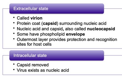
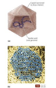
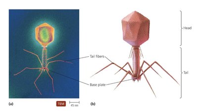
Virion Structure
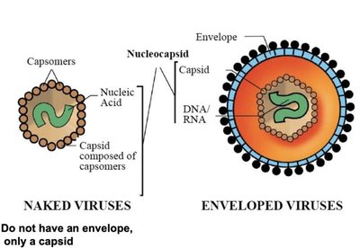
A virion is a complete virus particle, consisting of a nucleic acid, a protein coat (capsid), and sometimes a phospholipid envelope.
Capsid: Protects viral genome and aids in attachment to host cells; composed of protein subunits called capsomeres.
Nucleocapsid: Combination of nucleic acid and capsid.
Envelope: Some viruses acquire a phospholipid envelope from the host cell membrane, which contains glycoproteins important for host recognition.
Genetic Material
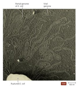
The type of nucleic acid (DNA or RNA) is the primary basis for virus classification. Viral genomes are much smaller than those of cells.
Can be single- or double-stranded, linear or circular.
Segmented genomes allow for genetic reassortment.
Hosts of Viruses
Viruses infect a wide range of hosts, including bacteria, plants, and animals. Host specificity is determined by the interaction between viral surface proteins and host cell receptors.
Some viruses are highly specific, infecting only certain cell types.
Generalist viruses can infect multiple cell types or species.

Capsid Morphology and Viral Shapes
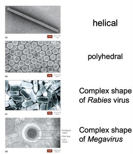
Viruses are classified by their capsid morphology and overall shape. There are three basic types:
Helical: Rod-shaped, with nucleic acid wound inside a helical capsid.
Polyhedral: Many-sided, often icosahedral.
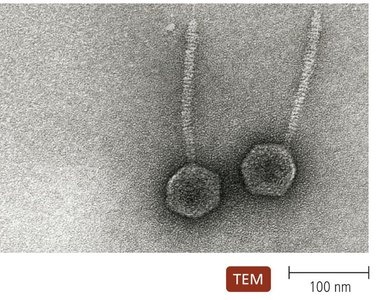
Complex: More intricate structures, such as bacteriophages.
The Viral Envelope
The envelope is acquired from the host cell during viral replication or release. It consists of a phospholipid bilayer and proteins, including virally coded glycoproteins (spikes).
Envelope proteins are crucial for host recognition.
Enveloped viruses are more fragile than naked viruses.
Classification of Viruses
Criteria for Classification
Viruses are classified based on:
Type of nucleic acid (DNA or RNA)
Presence or absence of an envelope
Shape and size
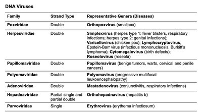
DNA Viruses
Family | Strand Type | Representative Genera (Diseases) |
|---|---|---|
Poxviridae | Double | Orthopoxvirus (smallpox) |
Herpesviridae | Double | Simplexvirus (herpes), Varicellovirus (chickenpox), Lymphocytovirus, Cytomegalovirus, Roseolovirus |
Papillomaviridae | Double | Papillomavirus (tumors, warts, cancers) |
Polyomaviridae | Double | Polyomavirus (leukoencephalopathy) |
Adenoviridae | Double | Mastadenovirus (conjunctivitis, respiratory infections) |
Hepadnaviridae | Partial single and partial double | Orthohepadnavirus (hepatitis B) |
Parvoviridae | Single | Erythrovirus (erythema infectiosum) |
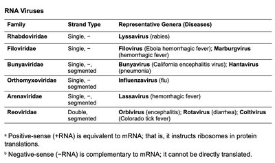
RNA Viruses
Family | Strand Type | Representative Genera (Diseases) |
|---|---|---|
Rhabdoviridae | Single, - | Lyssavirus (rabies) |
Filoviridae | Single, - | Filovirus (Ebola), Marburgvirus |
Bunyaviridae | Single, segmented | Bunyavirus (encephalitis), Hantavirus (pneumonia) |
Orthomyxoviridae | Single, segmented | Influenzavirus (flu) |
Arenaviridae | Single, segmented | Lassavirus (hemorrhagic fever) |
Reoviridae | Double, segmented | Orbivirus (encephalitis), Rotavirus (diarrhea), Coltivirus (Colorado tick fever) |
Viral Replication
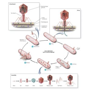
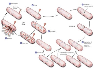
Lytic Replication Cycle
The lytic cycle results in the destruction of the host cell and the release of new virions. It consists of five stages:
Attachment
Entry
Synthesis
Assembly
Release
Lysogenic Replication Cycle
In lysogeny, the viral genome integrates into the host chromosome and remains dormant (prophage) until induced to enter the lytic cycle. Lysogenic conversion can alter the phenotype of the host bacterium. 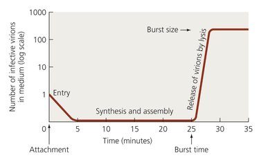
Replication of Animal Viruses
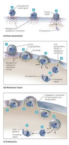
Animal viruses follow similar steps as bacteriophages but differ due to the presence of envelopes and the eukaryotic nature of host cells.
Attachment: Mediated by glycoprotein spikes or other molecules.
Entry and Uncoating: Can occur via direct penetration, membrane fusion, or endocytosis.
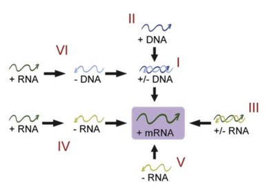
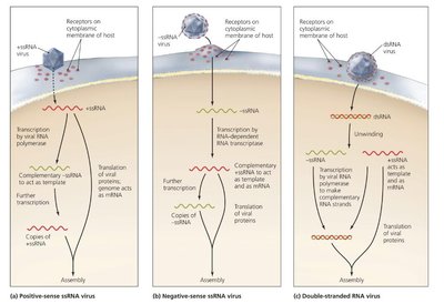
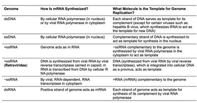
Synthesis Strategies
DNA Viruses: Often replicate in the nucleus; proteins synthesized in cytoplasm.
RNA Viruses: Replicate in cytoplasm; positive-sense RNA acts as mRNA, negative-sense RNA must be transcribed.
Retroviruses: Use reverse transcriptase to synthesize DNA from RNA.
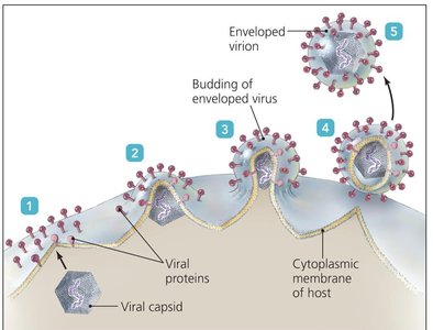
Assembly and Release
Most DNA viruses assemble in the nucleus; most RNA viruses in the cytoplasm.
Enveloped viruses are released by budding, which can result in persistent infections.
Naked viruses are released by exocytosis or lysis.
Latency
Some animal viruses remain dormant in host cells as latent viruses or proviruses. Incorporation into host DNA is permanent and may last for years.
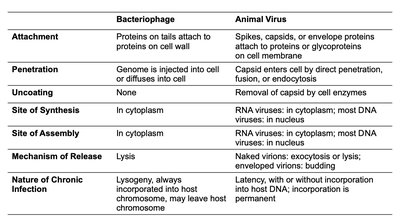
Comparison of Bacteriophage and Animal Virus Replication
Bacteriophage | Animal Virus | |
|---|---|---|
Attachment | Proteins on tails attach to proteins on cell wall | Spikes, capsids, or envelope proteins attach to proteins or glycoproteins on cell membrane |
Penetration | Genome is injected into cell or diffuses into cell | Capsid enters cell by direct penetration, fusion, or endocytosis |
Uncoating | None | Removal of capsid by cell enzymes |
Site of Synthesis | In cytoplasm | RNA viruses: in cytoplasm; most DNA viruses: in nucleus |
Site of Assembly | In cytoplasm | RNA viruses: in cytoplasm; most DNA viruses: in nucleus |
Mechanism of Release | Lysis | Naked virions: exocytosis or lysis; enveloped virions: budding |
Nature of Chronic Infection | Lysogeny, always incorporated into host chromosome, may leave host chromosome | Latency, with or without incorporation into host DNA; incorporation is permanent |
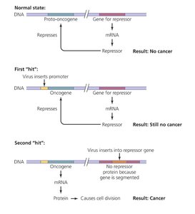
The Role of Viruses in Cancer
Oncogenes and Tumor Formation
Cell division is tightly regulated by genes. Viruses can disrupt this regulation by carrying oncogenes, promoting host oncogenes, or interfering with tumor repression, leading to neoplasia (tumor formation).
Malignant tumors are cancers; metastasis is the spread of cancer.
Environmental factors (UV light, radiation, carcinogens, viruses) can activate oncogenes.
Viruses and Human Cancers
Viruses may cause 20–25% of human cancers, including Burkitt’s lymphoma, Hodgkin’s disease, Kaposi’s sarcoma, and cervical cancer.
Culturing Viruses in the Laboratory
Methods of Culturing
Viruses cannot grow in standard media and must be cultured inside host cells. Three main methods:
Mature Organisms: Bacteria, plants, and animals; phages grown in bacteria produce plaques.
Embryonated Eggs: Commonly used for vaccine production; provide a nourishing environment.
Cell Cultures: Cells isolated and grown in media; includes diploid and continuous cell cultures.
Viroids and Prions
Viroids
Viroids are extremely small, circular pieces of ssRNA that are infectious and pathogenic in plants. They lack a capsid and do not code for proteins.
Viroid RNA adheres to complementary plant RNA, leading to degradation and disease.
Prions
Prions are proteinaceous infectious agents that cause neurodegenerative diseases.
Cellular PrP: Normal form with α-helices.
Prion PrP: Disease-causing form with β-pleated sheets; induces refolding of normal PrP.
Prion Diseases: Spongiform encephalopathies (BSE, scrapie, kuru, CWD, vCJD); transmitted by ingestion, transplantation, or contact.
No standard treatment; prions are resistant to normal sterilization, destroyed by incineration or autoclaving in sodium hydroxide.
Comparison of Viruses, Viroids, Prions, and Bacterial Cells
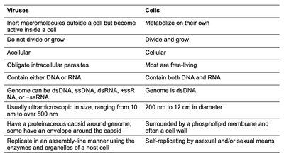
Viruses | Cells | |
|---|---|---|
Metabolism | Inert outside cell, active inside cell | Metabolize on their own |
Growth | Do not divide or grow | Divide and grow |
Cellularity | Acellular | Cellular |
Parasitism | Obligate intracellular parasites | Most are free-living |
Genetic Material | DNA or RNA | Both DNA and RNA |
Genome Type | dsDNA, ssDNA, dsRNA, +ssRNA, −ssRNA | dsDNA |
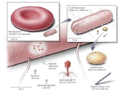
Size | 10 nm to 500 nm | 200 nm to 12 μm |
Structure | Capsid around genome; some have envelope | Phospholipid membrane, often cell wall |
Replication | Assembly-line manner using host cell | Self-replicating by asexual/sexual means |