 Back
BackViruses, Viroids, and Prions: Structure, Classification, and Pathogenicity
Study Guide - Smart Notes
 Tailored notes based on your materials, expanded with key definitions, examples, and context.
Viruses, Viroids, and Prions
Distinctive Features of Viruses
Viruses are unique infectious agents that differ fundamentally from cellular organisms. They are obligatory intracellular parasites, meaning they require living host cells to multiply. Viruses contain either DNA or RNA as their genetic material, but never both. Their structure includes a protein coat (capsid), and they lack ribosomes and an ATP-generating mechanism, relying entirely on the host cell's machinery for replication.
Obligatory intracellular parasites: Must invade host cells to reproduce.
Genetic material: DNA or RNA, single- or double-stranded, linear, circular, or segmented.
Protein coat: Protects genetic material and aids in host cell attachment.
No ribosomes or ATP-generating mechanism: Cannot synthesize proteins or generate energy independently.
Host Range
The host range of a virus refers to the spectrum of host cells it can infect. Most viruses are highly specific, infecting only certain cell types within a host. This specificity is determined by the presence of particular attachment sites on the host cell and compatible cellular factors.
Bacteriophages: Infect bacteria; receptor sites may be on cell wall, fimbriae, or flagella.
Animal viruses: Receptor sites are typically on the plasma membrane.
Virus Sizes
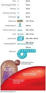
Viruses vary greatly in size, typically ranging from 20 nm to 300 nm. Their small size allows them to pass through bacteriological filters that retain bacteria.
Viral Structure
The virion is the complete, fully developed viral particle. Its structure includes:
Nucleic acid: DNA or RNA, single- or double-stranded, linear, circular, or segmented.
Capsid: Protein coat made of capsomeres (subunits).
Envelope: Lipid, protein, and carbohydrate coating present in some viruses.
Spikes: Projections from the outer surface, often used for attachment.
Capsid and Envelope
The capsid is composed of protein subunits called capsomeres. Some viruses possess an envelope external to the capsid, derived partly from the host cell's plasma membrane during viral budding. Spikes on the envelope may facilitate attachment to host cells.
General Morphology of Viruses
Viruses are classified by their morphology:
Helical viruses: Hollow, cylindrical capsid; e.g., rabies and Ebola viruses.
Polyhedral viruses: Many-sided, most commonly icosahedral; e.g., adenoviruses, poliovirus.
Enveloped viruses: Usually spherical.
Complex viruses: Complicated structures; e.g., bacteriophages.




Taxonomy and Classification of Viruses
Viruses are classified based on their genetic material and replication strategy. The Baltimore classification system divides viruses into seven groups (realms) according to their nucleic acid type and how mRNA is produced. Viral taxonomy uses the following conventions:
Genus: Ends in -virus
Family: Ends in -viridae
Order: Ends in -ales
Viral species: Group sharing genetic information and ecological niche

Growing Viruses in the Laboratory
Bacteriophages
Bacteriophages are grown in bacteria, forming plaques (clearings) on a lawn of bacteria on agar. Each plaque corresponds to a single virus and is quantified as plaque-forming units (PFU).

Animal Viruses
In living animals: Mice, rabbits, guinea pigs; some human viruses may not cause disease in animals.
In embryonated eggs: Virus injected into egg; growth signaled by changes or embryo death; used for vaccine production.

In cell cultures: Primary cell lines (from tissues), diploid cell lines (from embryos), and continuous cell lines (from cancerous cells, e.g., HeLa).
Cytopathic effect (CPE): Visible changes or deterioration in monolayer cells due to viral infection.


Viral Identification
Viruses are identified by:
Cytopathic effects: Observed in cell culture.
Serological tests: ELISA detects viruses by antibody reactions.
Nucleic acid tests: PCR amplifies viral genetic material for identification.
Viral Multiplication
To multiply, a virus must invade a host cell and commandeer its metabolic machinery. A single virion can produce thousands of progeny in one cell. The one-step growth curve illustrates the stages of viral replication.

Comparison of Bacteriophage and Animal Viral Multiplication
Stage | Bacteriophages | Animal Viruses |
|---|---|---|
Attachment | Tail fibers attach to cell wall proteins | Receptor sites are plasma membrane proteins and glycoproteins |
Entry | Viral DNA is injected into host cell | Capsid enters by receptor-mediated endocytosis or fusion |
Uncoating | Not required | Enzymatic removal of capsid proteins |
Biosynthesis | In cytoplasm | In nucleus (DNA viruses) or cytoplasm (RNA viruses) |
Chronic infection | Lysogeny | Latency; slow viral infections; cancer |
Release | Host cell is lysed | Enveloped viruses bud out; nonenveloped viruses rupture plasma membrane |

Multiplication of Animal Viruses
The process includes:
Attachment: Virus binds to cell membrane.
Entry: Via receptor-mediated endocytosis or fusion.
Uncoating: Viral nucleic acid separated from capsid by enzymes.
Biosynthesis: Production of viral nucleic acid and proteins.
Maturation: Assembly of nucleic acid and capsid proteins.
Release: By budding (enveloped viruses) or rupture (nonenveloped viruses).


Viruses and Cancer
Some viruses are oncogenic, meaning they can cause cancer. Viral-induced cancers may develop long after infection and are not contagious. Types include:
Sarcoma: Cancer of connective tissue.
Adenocarcinomas: Cancer of glandular epithelial tissue.
Transformation of Normal Cells Into Tumor Cells
Proto-oncogenes: Genes encoding proteins for normal cell growth; mutations convert them to oncogenes.
Oncogenic viruses: Integrate into host DNA, inducing tumors.
Transformed cells: Exhibit tumor-specific antigens and irregular shapes.
DNA Oncogenic Viruses
Adenoviridae
Herpesviridae: Epstein-Barr virus (Burkitt’s lymphoma)
Poxviridae
Papovaviridae: Human papillomavirus (HPV; cervical and anal cancer)
Hepadnaviridae: Hepatitis B virus
RNA Oncogenic Viruses
Retroviridae: Viral RNA transcribed to DNA (reverse transcriptase), integrates into host DNA. HTLV-1 and HTLV-2 cause adult T cell leukemia and lymphoma; FeLV causes feline leukemia.
Latent and Persistent Viral Infections
Viruses may establish latent or persistent infections:
Latent infections: Virus remains dormant in host cell; may reactivate (e.g., herpesviruses, cold sores, shingles).
Persistent infections: Virus is continuously released; often fatal (e.g., HIV/AIDS, hepatitis B, measles).

Disease | Primary Effect | Causative Virus |
|---|---|---|
Cold sores | Skin and mucous membrane lesions; genital lesions | HHV-1 and HHV-2 |
Leukemia | Increased white blood cell growth | HTLV-1 and -2 |
Shingles | Skin lesions | Varicellovirus (Herpesvirus) |
Cervical cancer | Increased cell growth | Human papillomavirus |
HIV/AIDS | Decreased CD4+ T cells | HIV-1 and -2 (Lentivirus) |
Liver cancer | Increased cell growth | Hepatitis B virus |
Persistent enterovirus infection | Mental deterioration associated with AIDS | Echoviruses |
Progressive encephalitis | Rapid mental deterioration | Rubella virus |
Subacute sclerosing panencephalitis (SSPE) | Mental deterioration | Measles virus |
Prions
Prions are infectious proteins, inherited and transmissible by ingestion, transplant, or surgical instruments. They cause spongiform encephalopathies such as "mad cow disease," Creutzfeldt-Jakob disease (CJD), and sheep scrapie. Disease results from the conversion of normal cellular prion protein (PrPC) into an infectious misfolded form (PrPSc), which accumulates in brain cells, forming plaques.
Viruses vs. Bacteria
Property | Bacteria | Viruses |
|---|---|---|
Intracellular Parasite | No (typical), Yes (Rickettsias/Chlamydias) | Yes |
Plasma Membrane | Yes | No |
Binary Fission | Yes | No |
Pass through Bacteriological Filters | No/Yes | Yes |
Possess Both DNA and RNA | Yes | No |
ATP-Generating Metabolism | Yes/No | No |
Ribosomes | Yes | No |
Sensitive to Antibiotics | Yes | No |
Sensitive to Interferon | No | Yes |
Summary
This chapter provides a comprehensive overview of viruses, viroids, and prions, including their structure, classification, replication, laboratory cultivation, pathogenicity, and comparison with bacteria. Understanding these agents is essential for microbiology students, as they play significant roles in infectious diseases, cancer, and neurodegenerative disorders.
