 Back
BackViruses, Viroids, and Prions: Structure, Classification, and Pathogenicity
Study Guide - Smart Notes
 Tailored notes based on your materials, expanded with key definitions, examples, and context.
Viruses, Viroids, and Prions
Viral Discovery and Historical Context
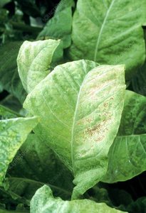
Viruses were first conceptualized as invisible infectious agents, described as contagium vivum fluidum (contagious living fluid), before they could be directly observed. The term "virus" comes from the Latin word for poison and was adopted in the 1930s to describe these filterable agents. Early studies, such as those by Adolf Mayer and Dmitri Ivanovsky, established that the causative agent of tobacco mosaic disease was smaller than bacteria and could pass through filters that retained bacteria.
Adolf Mayer (1886): Investigated tobacco mosaic disease and found it infectious but could not identify the microbe.
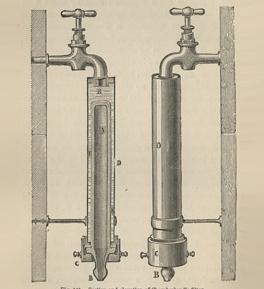
Dmitri Ivanovsky (1892): Used a Chamberland filter to show that the infectious agent could pass through bacteria-proof filters, suggesting it was smaller than bacteria or a toxin.
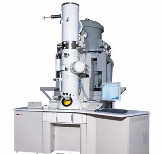
Electron Microscope (1930s): Enabled direct observation of viruses, allowing detailed study of their structure and replication.
Distinctive Features of Viruses
Viruses are obligate intracellular parasites that require living host cells to multiply. They contain either DNA or RNA (never both), are surrounded by a protein coat, and lack ribosomes and ATP-generating mechanisms. Viruses use the host cell's machinery for replication and protein synthesis.
Virion: A complete, fully developed, infectious viral particle outside a host cell.
Classification: Based on nucleic acid type (DNA or RNA) and protein coat structure.
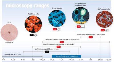
Viral Size and Visualization
Viruses are much smaller than bacteria and cannot be seen with light microscopes. The development of the electron microscope was crucial for visualizing viruses and understanding their morphology.
Viral Structure
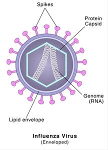
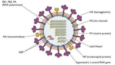
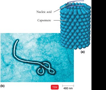
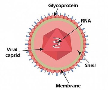
Viruses consist of a nucleic acid core (DNA or RNA) surrounded by a protein coat called a capsid, which is made up of protein subunits called capsomeres. Some viruses also have an outer lipid envelope derived from the host cell membrane, and may possess surface proteins called spikes for attachment and entry into host cells.
Nucleic Acid: Can be single- or double-stranded, linear, circular, or segmented. The genome size varies widely among viruses.
Capsid: Protein shell that protects the viral genome and aids in its delivery into host cells.
Envelope: Lipid membrane present in some viruses, acquired from the host cell during viral budding.
Spikes: Glycoprotein projections that facilitate attachment to host cells (e.g., influenza virus hemagglutinin and neuraminidase).
Virus Morphology and Categories
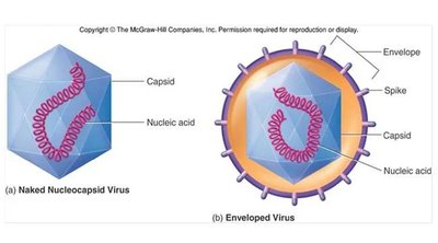
Viruses are classified based on their morphology and the presence or absence of an envelope:
Naked (Non-enveloped) Viruses: Consist only of nucleic acid and capsid.
Enveloped Viruses: Have a nucleic acid-capsid core surrounded by a lipid envelope.
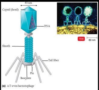
Complex Viruses: Exhibit complicated structures, such as bacteriophages with heads, tails, and fibers.
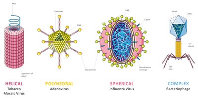
Common Virus Shapes
Helical Viruses: Rod-shaped, with nucleic acid inside a helical capsid (e.g., rabies, Ebola).
Polyhedral Viruses: Many-sided, typically icosahedral (e.g., poliovirus).
Enveloped Viruses: Spherical, with helical or polyhedral nucleocapsids inside a lipid envelope (e.g., influenza, Marburgvirus).
Complex Viruses: Bacteriophages with additional structures for infecting bacteria.
Virus Classification
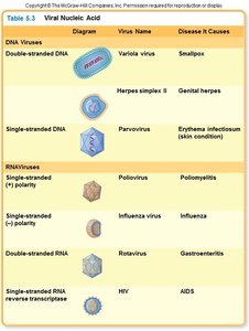
Viruses are classified by their nucleic acid type, capsid symmetry, presence or absence of an envelope, and host range. The table below summarizes medically important viruses by nucleic acid type and structure.
Genome Type | Capsid Shape | Envelope | Example Virus |
|---|---|---|---|
dsDNA | Icosahedral | Naked/Enveloped | Herpesvirus |
ssRNA (+) | Icosahedral | Naked/Enveloped | Poliovirus, Influenza |
ssRNA (-) | Helical | Enveloped | Rabies, Ebola |
dsRNA | Icosahedral | Naked | Rotavirus |
Retrovirus (ssRNA-RT) | Icosahedral | Enveloped | HIV |
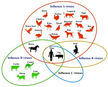
Host Range and Transmission
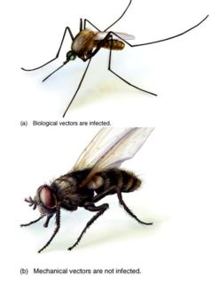
The host range of a virus is the spectrum of host cells it can infect, determined by specific attachment sites and cellular factors. Viruses can infect bacteria (bacteriophages), plants, or animals. Transmission occurs via direct contact, indirect contact with fomites, or vectors (e.g., mosquitoes, ticks, flies). Vectors may transmit viruses biologically (internal) or mechanically (external).
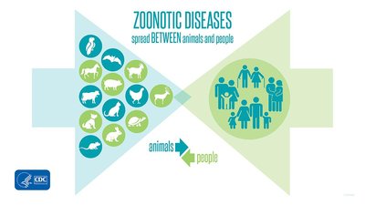
Zoonoses: Diseases transmitted from animals to humans (e.g., avian influenza).
Reverse Zoonoses: Transmission from humans to animals.

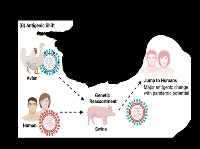
Genetic Variation: Antigenic Drift and Shift
Viruses, especially RNA viruses, undergo genetic changes that impact their epidemiology and immune evasion:
Antigenic Drift: Accumulation of small mutations due to lack of proofreading during replication, resulting in minor antigenic changes and new viral variants.
Antigenic Shift: Reassortment of RNA segments from different viruses, leading to major antigenic changes and the emergence of new subtypes with pandemic potential.
Viral Multiplication in Animal Cells
Viral replication in animal cells follows a series of steps:
Attachment: Virus binds to host cell membrane receptors.
Entry: Virus enters the cell via receptor-mediated endocytosis or membrane fusion.
Uncoating: Viral genome is released from the capsid by host or viral enzymes.
Biosynthesis: Viral nucleic acid and proteins are synthesized using host machinery.
Maturation: Assembly of viral components into new virions.
Release: New virions exit the cell by budding (enveloped viruses) or cell lysis (naked viruses).
Viruses and Cancer
Some viruses are oncogenic, meaning they can cause cancer by integrating their genetic material into the host genome and disrupting normal cell regulation. This process may occur long after the initial infection and is not contagious. Oncogenic transformation involves the activation of proto-oncogenes into oncogenes, leading to uncontrolled cell growth and tumor formation.
Examples: Human papillomavirus (HPV) and hepatitis B virus (HBV) are associated with cervical and liver cancers, respectively.
Latent and Persistent Viral Infections
Some viruses can establish latent infections, remaining dormant within host cells and reactivating under certain conditions (e.g., herpesviruses). Persistent viral infections involve continuous, long-term production of virus, often leading to chronic disease and sometimes fatal outcomes.
Latent Infection: Virus remains inactive but can reactivate (e.g., herpes simplex virus).
Persistent Infection: Virus is continuously produced over a long period (e.g., HIV, hepatitis B).
Viroids and Prions
Viroids are infectious RNA molecules that lack a protein coat and primarily infect plants. Prions are infectious proteins that cause neurodegenerative diseases in animals and humans. Prion diseases are inherited or acquired through ingestion, transplantation, or contaminated surgical instruments, and are characterized by spongiform encephalopathies (e.g., mad cow disease, Creutzfeldt-Jakob disease).
Prion Diseases: Mad cow disease, Creutzfeldt-Jakob disease, Gerstmann-Sträussler-Scheinker syndrome, fatal familial insomnia, sheep scrapie, and kuru.
