 Back
BackViruses, Viroids, and Prions: Structure, Classification, and Multiplication
Study Guide - Smart Notes
 Tailored notes based on your materials, expanded with key definitions, examples, and context.
Viruses, Viroids, and Prions
General Characteristics of Viruses
Viruses are unique infectious agents that differ significantly from bacteria and other microorganisms. They are obligate intracellular parasites, meaning they require living host cells to multiply. Viruses contain either DNA or RNA as their genetic material, but never both. They lack ribosomes and an ATP-generating mechanism, making them entirely dependent on host cellular machinery for replication.
Obligate intracellular parasites: Cannot reproduce outside a living cell.
Genetic material: DNA or RNA, single- or double-stranded, linear or circular.
Protein coat (capsid): Protects the genetic material.
No ribosomes or ATP generation: Cannot synthesize proteins or generate energy independently.
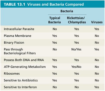
Table Purpose: This table compares the fundamental properties of typical bacteria, rickettsias/chlamydias, and viruses, highlighting differences in structure, metabolism, and sensitivity to antibiotics and interferon.
Host Range and Size
The host range of a virus refers to the spectrum of host cells it can infect. Most viruses are highly specific, infecting only certain cell types within a single host species. This specificity is determined by the presence of compatible attachment sites and cellular factors. Bacteriophages are viruses that infect bacteria. Viruses are much smaller than most cells, typically ranging from 20 nm to 1000 nm in length.
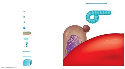
Image Purpose: This image visually compares the sizes of various viruses, bacteria, and eukaryotic cells, emphasizing the small size of viruses relative to other biological entities.
Viral Structure
Virion Structure
A virion is a complete, fully developed viral particle capable of causing infection. The main components of a virion include:
Nucleic acid: DNA or RNA, which carries the genetic information.
Capsid: Protein coat made of subunits called capsomeres.
Envelope (in some viruses): Lipid, protein, and carbohydrate layer derived from the host cell membrane.
Spikes: Protein projections that aid in attachment to host cells.
Types of Viral Morphology
Helical viruses: Hollow, cylindrical capsid.
Polyhedral viruses: Many-sided, often icosahedral.
Enveloped viruses: Surrounded by a lipid envelope.
Complex viruses: Complicated structures, such as bacteriophages.
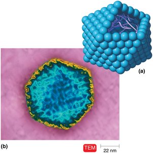
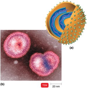
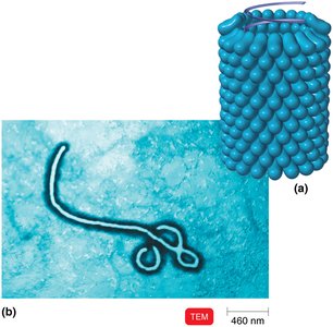
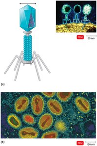
Image Purpose: These images illustrate the structural diversity of viruses, including polyhedral, helical, enveloped, and complex forms.
Taxonomy of Viruses
Classification and Nomenclature
Viruses are classified based on their genetic material, structure, and host range. The taxonomy of viruses includes:
Genus names: End in -virus (e.g., Lentivirus).
Family names: End in -viridae (e.g., Retroviridae).
Order names: End in -ales.
Viral species: Group of viruses sharing genetic information and ecological niche.
Subspecies: Designated by numbers (e.g., HIV-1, HIV-2).
Example: Herpesviridae (family), Herpesvirus (genus), Human herpes virus (species), HHV-1, HHV-2, HHV-3 (subspecies).
Isolation, Cultivation, and Identification of Viruses
Growing Bacteriophages
Bacteriophages are grown in bacterial cultures. When bacteriophages infect bacteria on an agar plate, they form clear zones called plaques, each representing a single virus. The number of plaques can be quantified as plaque-forming units (PFU).
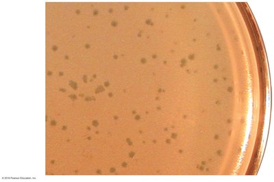
Image Purpose: This image shows plaques on a bacterial lawn, which are used to quantify bacteriophage concentration.
Growing Animal Viruses
In living animals: Used for viruses that do not grow well in vitro.
In embryonated eggs: Virus is injected into various egg compartments; growth is detected by embryo changes or death.
In cell cultures: Tissues are enzymatically treated to separate cells, which are then grown in culture. Viral infection is detected by the cytopathic effect (CPE), or cell deterioration.
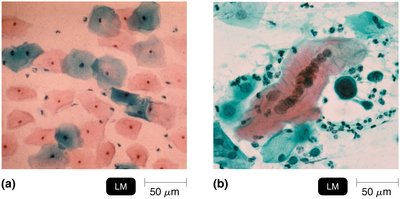
Image Purpose: This image demonstrates the cytopathic effect, a key method for identifying viral infection in cultured cells.
Viral Identification
Cytopathic effects: Observable changes in host cells.
Serological tests: Detection of viral antigens or antibodies (e.g., Western blotting).
Nucleic acid analysis: Techniques such as RFLPs and PCR.
Viral Multiplication
One-Step Growth Curve
The replication of viruses follows a one-step growth curve, with a latent (eclipse) period followed by a sharp increase in virion numbers as host cells are lysed and new virions are released.
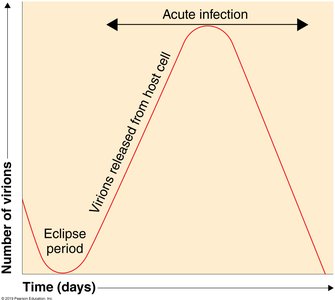
Image Purpose: This graph illustrates the stages of viral replication, including the eclipse period and acute infection phase.
Multiplication of Bacteriophages
Lytic cycle: Phage causes lysis and death of the host cell.
Lysogenic cycle: Phage DNA integrates into host DNA as a prophage, replicating with the host genome and potentially conferring new properties (phage conversion).

Image Purpose: This diagram shows the steps of the lysogenic and lytic cycles in bacteriophage lambda.
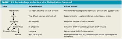
Comparison: Bacteriophage vs. Animal Virus Multiplication
Stage | Bacteriophages | Animal Viruses |
|---|---|---|
Attachment | Tail fibers attach to cell wall proteins | Attachment sites are plasma membrane proteins/glycoproteins |
Entry | Viral DNA injected into host cell | Capsid enters by endocytosis or fusion |
Uncoating | Not required | Enzymatic removal of capsid proteins |
Biosynthesis | In cytoplasm | In nucleus (DNA viruses) or cytoplasm (RNA viruses) |
Chronic Infection | Lysogeny | Latency, slow viral infections, cancer |
Release | Host cell is lysed | Enveloped viruses bud out; nonenveloped viruses rupture plasma membrane |
Multiplication of Animal Viruses
Attachment: Virus binds to specific receptors on the cell surface.
Entry: By receptor-mediated endocytosis or fusion.
Uncoating: Separation of viral nucleic acid from the capsid.
Biosynthesis: Synthesis of viral nucleic acids and proteins.
Maturation: Assembly of viral components into new virions.
Release: By budding (enveloped viruses) or rupture (nonenveloped viruses).
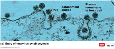
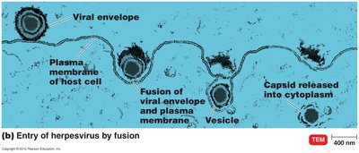
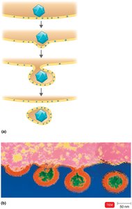
Image Purpose: These images illustrate the mechanisms of viral entry (pinocytosis and fusion) and release (budding) in animal cells.
Biosynthesis of DNA and RNA Viruses
DNA viruses: Replicate DNA in the nucleus using viral enzymes; synthesize capsid proteins in the cytoplasm.
RNA viruses: Replicate in the cytoplasm; mechanisms vary by virus type.
Retroviruses: Use reverse transcriptase to synthesize DNA from RNA, which integrates into the host genome as a provirus.
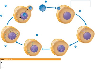
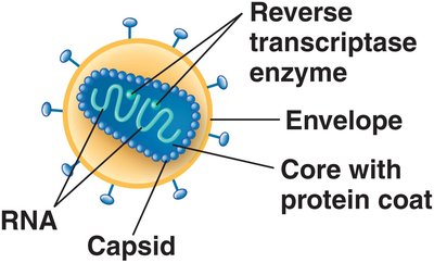
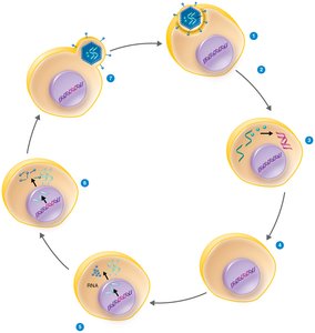
Image Purpose: These images show the replication cycles of DNA viruses and retroviruses, highlighting key steps such as reverse transcription and integration.
Viruses and Cancer
Oncogenes and Transformation
Oncogenes are genes that can transform normal cells into cancerous cells. Oncogenic viruses integrate their genetic material into the host genome, potentially activating oncogenes and inducing tumor formation. Transformed cells often display unique antigens and altered growth properties.
DNA oncogenic viruses: Adenoviridae, Herpesviridae (e.g., Epstein-Barr virus), Papovaviridae (e.g., human papillomavirus), Hepadnaviridae (e.g., hepatitis B virus).
RNA oncogenic viruses: Retroviridae (e.g., HTLV-1, HTLV-2).
Latent and Persistent Viral Infections
Definitions and Examples
Latent viral infections: Virus remains dormant in host cells and may reactivate (e.g., cold sores, shingles).
Persistent viral infections: Virus is continuously present and often fatal (e.g., subacute sclerosing panencephalitis caused by measles virus).
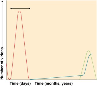
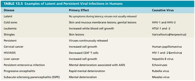
Image Purpose: The graph and table illustrate the differences between acute, latent, and persistent infections, and provide examples of diseases caused by each type.
Summary Table: Key Differences Between Viruses and Bacteria
Feature | Typical Bacteria | Rickettsias/Chlamydias | Viruses |
|---|---|---|---|
Intracellular Parasite | No | Yes | Yes |
Plasma Membrane | Yes | Yes | No |
Binary Fission | Yes | Yes | No |
Pass through Bacteriological Filters | No | No/Yes | Yes |
Possess Both DNA and RNA | Yes | Yes | No |
ATP-Generating Metabolism | Yes | No | No |
Ribosomes | Yes | Yes | No |
Sensitive to Antibiotics | Yes | Yes | No |
Sensitive to Interferon | No | No | Yes |
Table Purpose: This table summarizes the distinguishing features of viruses compared to typical bacteria and rickettsias/chlamydias.
