 Back
Back13 Viruses, Viroids, and Prions: Structure, Classification, and Replication
Study Guide - Smart Notes
 Tailored notes based on your materials, expanded with key definitions, examples, and context.
Viruses, Viroids, and Prions
General Characteristics of Viruses
Viruses are unique infectious agents that differ significantly from bacteria and other microorganisms. They are obligate intracellular parasites, meaning they require living host cells to multiply. Viruses contain a single type of nucleic acid (either DNA or RNA), a protein coat (capsid), and sometimes an envelope derived from the host cell membrane. They lack ribosomes and ATP-generating mechanisms, making them entirely dependent on host cellular machinery for replication.
Obligate intracellular parasites: Cannot reproduce outside a living cell.
Genome: Contains either DNA or RNA, never both.
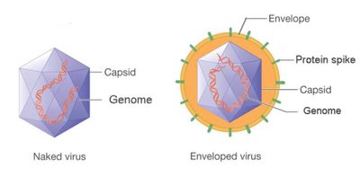
Capsid: Protein coat that protects the nucleic acid.
Envelope: Lipid, protein, and carbohydrate layer present in some viruses.
No ribosomes or ATP-generating mechanisms: Cannot synthesize proteins or generate energy independently.
Viruses and Bacteria Compared
Viruses differ from bacteria in several fundamental ways. The following table summarizes key differences:
Feature | Typical Bacteria | Rickettsias/Chlamydias | Viruses |
|---|---|---|---|
Intracellular Parasite | No | Yes | Yes |
Plasma Membrane | Yes | Yes | No |
Binary Fission | Yes | Yes | No |
Pass through Bacteriological Filters | No | No/Yes | Yes |
Possess Both DNA and RNA | Yes | Yes | No |
ATP-Generating Metabolism | Yes | Yes/No | No |
Ribosomes | Yes | Yes | No |
Sensitive to Antibiotics | Yes | Yes | No |
Sensitive to Interferon | No | No | Yes |
Host Range
The host range of a virus is the spectrum of host cells it can infect. Most viruses infect only specific types of cells in one host, determined by specific attachment sites and cellular factors.
Attachment sites: Viral surface proteins interact with specific host cell receptors.
Cellular factors: Host cell machinery must support viral replication.
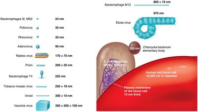
Virus Sizes
Viruses are much smaller than bacteria, typically ranging from 20 to 1000 nanometers in length. Their small size allowed researchers to detect their presence before the invention of the electron microscope, as they could pass through filters that retained bacteria.
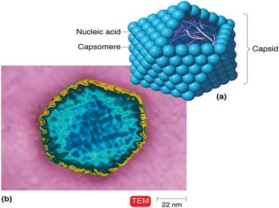
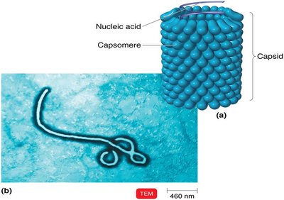
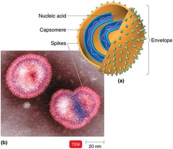
Viral Structure and Morphology
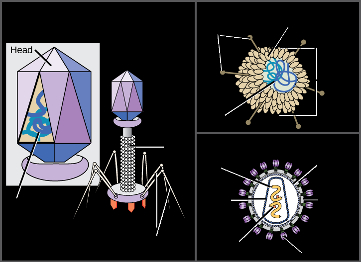
Virion Structure
A virion is a complete, fully developed viral particle. Its main components include:
Nucleic acid: DNA or RNA, single- or double-stranded, linear or circular.
Capsid: Protein coat made of capsomeres (subunits).
Envelope: Lipid, protein, and carbohydrate coating present in some viruses.
Spikes: Glycoprotein projections from the outer surface, important for attachment.
General Morphology
Viruses exhibit several morphological types:
Helical viruses: Hollow, cylindrical capsid (e.g., Ebola virus).
Polyhedral viruses: Many-sided, often icosahedral (e.g., adenovirus).
Enveloped viruses: Surrounded by a lipid envelope (e.g., influenza virus).
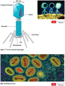
Complex viruses: Complicated structures, such as bacteriophages with a head, tail, and tail fibers.
Taxonomy of Viruses
Classification and Nomenclature
Viruses are classified based on their genetic material, structure, and host range. The taxonomy uses the following conventions:
Genus names: End in -virus
Family names: End in -viridae
Order names: End in -ales
Viral species: Group of viruses sharing the same genetic information and ecological niche (host)
Subspecies: Designated by a number
Examples of Virus Families
Family | Genus | Genome Type | Clinical Features |
|---|---|---|---|
Adenoviridae | Mastadenovirus | dsDNA, nonenveloped | Respiratory infections, tumors in animals |
Poxviridae | Orthopoxvirus | dsDNA, enveloped | Smallpox, cowpox |
Herpesviridae | Simplexvirus, Varicellovirus | dsDNA, enveloped | Cold sores, chickenpox, mononucleosis |
Picornaviridae | Enterovirus, Rhinovirus | ssRNA (+), nonenveloped | Polio, common cold, hepatitis A |
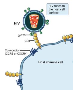
Retroviridae | Lentivirus (HIV) | ssRNA-RT, enveloped | AIDS |
Isolation, Cultivation, and Identification of Viruses
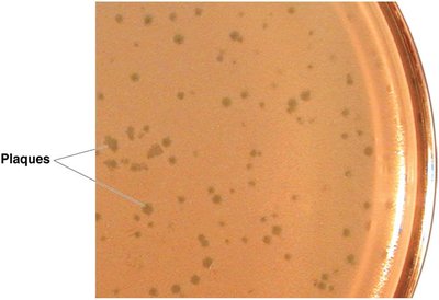
Growing Bacteriophages
Bacteriophages are grown in bacteria. They form plaques (clearings) on a lawn of bacteria on agar plates. Each plaque corresponds to a single virus and can be quantified as plaque-forming units (PFU).
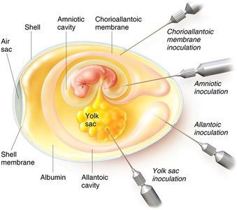
Growing Animal Viruses
In living animals: Used to study immune responses.
In embryonated eggs: Virus injected into egg; growth indicated by embryo changes or death.
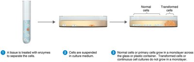
In cell cultures: Tissues treated with enzymes to separate cells; virally infected cells show cytopathic effect (CPE). Continuous cell lines are immortal and preferred for routine culture.
Viral Identification
Cytopathic effects: Observable changes in host cells due to viral infection.
Serological tests: Detection of viral proteins using antibodies (e.g., Western blotting).
Nucleic acid tests: RFLPs and PCR for genome analysis and amplification.
Viral Multiplication
Lytic and Lysogenic Cycles in Bacteriophages
Bacteriophages can undergo two types of replication cycles:
Lytic cycle: Phage causes lysis and death of the host cell.
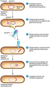
Lysogenic cycle: Phage DNA integrates into host DNA as a prophage and replicates with the host cell, potentially conferring new properties (phage conversion).

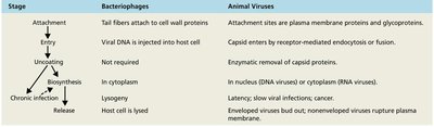
Steps of the Lytic Cycle (T-Even Bacteriophages)
Attachment: Phage attaches to host cell.
Penetration: Phage injects DNA into host.
Biosynthesis: Phage DNA and proteins are synthesized.
Maturation: Assembly of phage particles.
Release: Host cell lyses, releasing new phages.

Lysogenic Cycle (Bacteriophage Lambda λ)
Phage DNA integrates into host genome as a prophage.
Prophage is replicated with host DNA and can confer new traits (e.g., toxin production).
Specialized transduction: Only specific bacterial genes near the integration site are transferred by the phage.
Multiplication of Animal Viruses
Animal viruses follow a similar but distinct replication process:
Attachment: Virus binds to cell membrane receptors.
Entry: By receptor-mediated endocytosis or fusion.
Uncoating: Viral or host enzymes remove capsid.
Biosynthesis: Synthesis of viral nucleic acid and proteins.
Maturation: Assembly of viral components.
Release: By budding (enveloped viruses) or rupture (nonenveloped viruses).
Biosynthesis of DNA and RNA Viruses
DNA Viruses
Replicate DNA in the nucleus using viral enzymes.
Synthesize capsid proteins in the cytoplasm using host enzymes.
RNA Viruses
Replicate in the cytoplasm using RNA-dependent RNA polymerase.
+ssRNA (sense): Serves directly as mRNA.
-ssRNA (antisense): Must be transcribed to +ssRNA before translation.
dsRNA: Both strands present; replication involves synthesis of mRNA from the template.
Retroviruses
Single-stranded RNA viruses that produce DNA using reverse transcriptase.
Viral DNA integrates into host genome as a provirus.
Viruses and Cancer
Oncogenes and Transformation
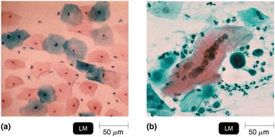
Some viruses can cause cancer by integrating their genetic material into the host genome, leading to transformation of normal cells into tumor cells. These viruses are called oncogenic viruses.
Oncogenes: Genes that can transform a normal cell into a cancerous cell.
Transformed cells: Exhibit uncontrolled growth and express tumor-specific antigens.
Examples of Oncogenic Viruses
DNA oncogenic viruses: Adenoviridae, Herpesviridae (Epstein-Barr virus), Papovaviridae (HPV), Hepadnaviridae (Hepatitis B virus).
RNA oncogenic viruses: Retroviridae (HTLV-1, HTLV-2).
Latent and Persistent Viral Infections
Latent Viral Infections
Virus remains dormant in host cells for long periods.
May reactivate due to changes in immunity (e.g., cold sores, shingles).
Persistent Viral Infections
Virus is continuously released over a long period, often leading to fatal outcomes (e.g., HIV/AIDS, liver cancer from hepatitis B).
Plant Viruses and Viroids
Plant Viruses
Enter plant cells through wounds or via insect vectors.
Protected by the plant cell wall, which limits infection.
Viroids and Virusoids
Viroids: Short pieces of naked RNA that cause plant diseases (e.g., potato spindle tuber disease).
Virusoids: Viroids enclosed in a protein coat; require coinfection with a virus to cause disease.
Prions
Prion Diseases
Prions are infectious proteins that cause neurodegenerative diseases. They are inherited or transmissible by ingestion, transplant, or surgical instruments. Prion diseases include:
Creutzfeldt-Jakob disease (CJD)
Kuru
Gerstmann-Sträussler-Scheinker syndrome
Fatal familial insomnia
Sheep scrapie
Prions exist in two forms: PrPC (normal) and PrPSc (misfolded, infectious). PrPSc accumulates in brain cells, forming plaques and causing disease.
