Back
Viruses, Viroids, and Prions: Structure, Classification, and Pathogenicity
Study Guide - Smart Notes
 Tailored notes based on your materials, expanded with key definitions, examples, and context.
Viruses, Viroids, and Prions
General Characteristics of Viruses
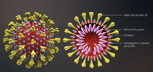
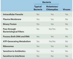
Viruses are obligatory intracellular parasites that require living host cells to multiply. Unlike bacteria, they do not possess ribosomes or an ATP-generating mechanism. Viruses contain either DNA or RNA (never both), and are surrounded by a protein coat called a capsid. Some viruses also have an envelope derived from the host cell membrane, and may have surface projections called spikes that aid in attachment to host cells.
Host Range: The spectrum of host cells a virus can infect is determined by specific attachment sites and cellular factors. Most viruses infect only specific types of cells in one host species.
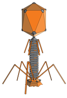
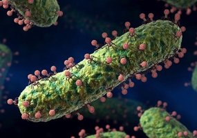
Bacteriophages: Viruses that infect bacteria are called bacteriophages.
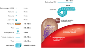
Size: Viruses range from 20 nm to 1000 nm in length, much smaller than most bacteria.
Example: Hepatitis viruses specifically infect liver cells, while SARS-CoV-2 infects respiratory tract cells.
Virus Structure
A virion is a complete, fully developed viral particle. The main components include:
Nucleic Acid: DNA or RNA, which may be single- or double-stranded, linear or circular.
Capsid: Protein coat made of subunits called capsomeres.
Envelope: Lipid, protein, and carbohydrate coating present in some viruses.
Spikes: Projections from the outer surface, important for attachment to host cells.
General Morphology of Viruses
Helical Viruses: Hollow, cylindrical capsid (e.g., tobacco mosaic virus).
Polyhedral Viruses: Many-sided (e.g., adenovirus).
Enveloped Viruses: Surrounded by a lipid envelope (e.g., influenza virus).
Complex Viruses: Complicated structures, such as bacteriophages.
Virus Taxonomy
Classification and Nomenclature
Viruses are classified based on their genetic material, morphology, and host range. The taxonomy includes:
Genus names: End in -virus
Family names: End in -viridae
Order names: End in -ales
Viral species: Group of viruses sharing the same genetic information and ecological niche (host)
Subspecies: Designated by a number
Isolation, Cultivation, and Identification of Viruses
Methods of Cultivation
Bacteriophages: Grown in bacteria, forming plaques (clearings) on a lawn of bacteria on agar. Each plaque corresponds to a single virus and can be quantified as plaque-forming units (PFU).
In Living Animals: Some viruses are grown in laboratory animals.
In Embryonated Eggs: Viruses are injected into eggs; viral growth is indicated by changes or death of the embryo.
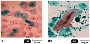
In Cell Cultures: Tissues are treated with enzymes to separate cells, and virally infected cells are detected by cytopathic effects (CPE). Continuous cell lines are often used for virus cultivation.
Viral Identification Techniques
Cytopathic Effects: Observable changes in host cells due to viral infection.
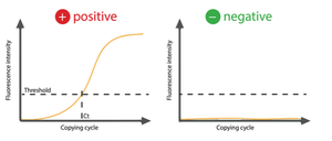
Nucleic Acid Tests: Detection of viral DNA or RNA using polymerase chain reaction (PCR). Real-time PCR allows rapid detection of viral nucleic acids.

Antigen Tests: Detect viral proteins using antibodies; color change indicates presence of virus.
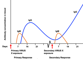
Antibody Tests: Detect host antibodies (IgM and IgG) against viral components, indicating exposure or immune response.
Viral Multiplication
One-Step Growth Curve
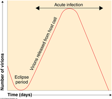
Viral replication follows a one-step growth curve, with an eclipse period (no detectable virions) followed by a sharp increase in virion numbers as they are released from host cells.
Multiplication of Bacteriophages
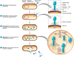
Lytic Cycle: Phage causes lysis and death of the host cell.
Lysogenic Cycle: Phage DNA is incorporated into the host DNA as a prophage, which can later enter the lytic cycle.
Phage Conversion: Host cell exhibits new properties due to prophage genes.
Specialized Transduction: Specific bacterial genes are transferred to another bacterium via a phage.
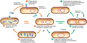
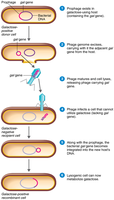
Comparison: Bacteriophage vs. Animal Virus Multiplication
Stage | Bacteriophages | Animal Viruses |
|---|---|---|
Attachment | Tail fibers attach to cell wall proteins | Attachment sites are plasma membrane proteins and glycoproteins |
Entry | Viral DNA is injected into host cell | Capsid enters by receptor-mediated endocytosis or fusion |
Uncoating | Not required | Enzymatic removal of capsid proteins |
Biosynthesis | In cytoplasm | In nucleus (DNA viruses) or cytoplasm (RNA viruses) |
Chronic Infection | Lysogeny | Latency, slow viral infections, cancer |
Release | Host cell is lysed | Enveloped viruses bud out; nonenveloped viruses rupture plasma membrane |

Animal Virus Classes and Examples
DNA Viruses
Adenoviridae: Double-stranded DNA, nonenveloped; causes respiratory infections and tumors.
Poxviridae: Double-stranded DNA, enveloped; causes skin lesions (e.g., smallpox).
Herpesviridae: Double-stranded DNA, enveloped; includes herpes simplex virus, varicella-zoster virus, Epstein-Barr virus, cytomegalovirus, and others.
Papovaviridae: Double-stranded DNA, nonenveloped; includes human papillomavirus (HPV), which can cause warts and cancer.
Hepadnaviridae: Double-stranded DNA, enveloped; includes hepatitis B virus, which uses reverse transcriptase.
RNA Viruses
Picornaviridae: Single-stranded RNA, + strand, nonenveloped; includes poliovirus, rhinovirus, hepatitis A virus.
Coronaviridae: Single-stranded RNA, + strand, enveloped; includes SARS, SARS-CoV-2.
Flaviviridae: Single-stranded RNA, + strand, enveloped; includes West Nile virus, hepatitis C, Zika virus.
Togaviridae: Single-stranded RNA, + strand, enveloped; includes rubella virus, chikungunya virus.
Rhabdoviridae: Single-stranded RNA, − strand; includes rabies virus.
Reoviridae: Double-stranded RNA, nonenveloped; includes rotavirus.
Retroviridae: Single-stranded RNA, produces DNA using reverse transcriptase; includes HIV.
Viruses and Cancer
Oncogenes and Transformation
Some viruses can cause cancer by integrating their genetic material into the host genome, leading to transformation of normal cells into tumor cells. Oncogenes are mutated proto-oncogenes that drive uncontrolled cell growth. Oncogenic viruses may induce tumors by expressing viral oncogenes or by disrupting host regulatory genes.
DNA Oncogenic Viruses: Adenoviridae, Herpesviridae (e.g., Epstein-Barr virus), Papovaviridae (HPV), Hepadnaviridae (hepatitis B virus).
RNA Oncogenic Viruses: Retroviridae (e.g., HTLV-1, HTLV-2).

Latent and Persistent Viral Infections
Definitions and Examples
Latent Viral Infections: Virus remains dormant in host cells and may reactivate (e.g., herpes simplex virus causing cold sores, varicella-zoster virus causing shingles).
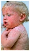
Persistent Viral Infections: Virus is continuously present and replicates at low levels, often leading to progressive disease (e.g., subacute sclerosing panencephalitis from measles virus).
Prions
Proteinaceous Infectious Particles
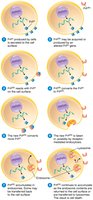
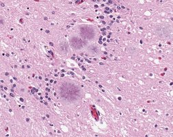
Prions are infectious proteins that lack nucleic acids. They are inherited and transmissible by ingestion, transplantation, or surgical instruments. Prions cause spongiform encephalopathies, a group of fatal neurodegenerative diseases characterized by sponge-like changes in brain tissue.
Examples: Mad cow disease (bovine spongiform encephalopathy), Creutzfeldt-Jakob disease (CJD), Gerstmann-Sträussler-Scheinker syndrome, fatal familial insomnia, sheep scrapie.