Back
Viruses, Viroids, and Prions: Structure, Classification, and Pathogenesis
Study Guide - Smart Notes
 Tailored notes based on your materials, expanded with key definitions, examples, and context.
Viruses, Viroids, and Prions
Distinctive Features of Viruses
Viruses are unique infectious agents that differ fundamentally from cellular organisms. They are obligatory intracellular parasites, meaning they require living host cells to multiply. Viruses contain either DNA or RNA as their genetic material, but never both, and possess a protein coat called a capsid. They lack ribosomes and an ATP-generating mechanism, relying entirely on the host cell's machinery for replication.
Obligatory intracellular parasites: Must invade host cells to reproduce.
Genetic material: DNA or RNA, single- or double-stranded, linear, circular, or segmented.
Protein coat: Capsid made of capsomeres.
No ribosomes or ATP generation: Cannot synthesize proteins or energy independently.
Host Range
The host range of a virus refers to the spectrum of host cells it can infect. Most viruses are highly specific, infecting only certain cell types within a host. This specificity is determined by the presence of particular attachment sites on the host cell and compatible cellular factors.
Bacteriophages: Infect bacteria; receptor sites may be on cell wall, fimbriae, or flagella.
Animal viruses: Receptor sites are typically on the plasma membrane.
Virus Sizes
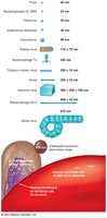
Viruses vary greatly in size, generally much smaller than bacteria. Their size influences their detection and classification.
Viral Structure
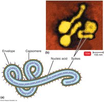
A virion is a complete, fully developed viral particle. The structure includes:
Nucleic acid: DNA or RNA, single- or double-stranded, linear, circular, or segmented.
Capsid: Protein coat made of capsomeres.
Envelope: Lipid, protein, and carbohydrate coating present in some viruses, often derived from the host cell membrane.
Spikes: Projections from the envelope, used for attachment.
General Morphology of Viruses
Viruses are classified by their morphology:
Helical viruses: Hollow, cylindrical capsid (e.g., rabies, Ebola).
Polyhedral viruses: Many-sided, often icosahedral (e.g., adenovirus, poliovirus).
Enveloped viruses: Spherical, with an envelope.
Complex viruses: Complicated structures (e.g., bacteriophage).
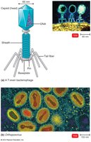
Taxonomy of Viruses
Viruses are classified using the Baltimore classification system, which is based on the type of nucleic acid and the method of mRNA production. Viral taxonomy includes:
Genus: Ends in -virus
Family: Ends in -viridae
Order: Ends in -ales
Viral species: Group sharing genetic information and ecological niche
Virus Cultivation and Identification
Growing Bacteriophages in the Laboratory
Bacteriophages are grown in bacteria, forming plaques—clearings on a lawn of bacteria. Each plaque corresponds to a single virus and is measured as plaque-forming units (PFU).
Growing Animal Viruses in the Laboratory
Animal viruses can be cultivated in living animals, embryonated eggs, or cell cultures. Cell cultures are the most common method, involving primary, diploid, or continuous cell lines.
Primary cell lines: Derived from tissues, form monolayers, die out after a few generations.
Diploid cell lines: Derived from human embryos, maintained for about 100 generations.
Continuous cell lines: Derived from cancerous cells, maintained indefinitely (e.g., HeLa cells).
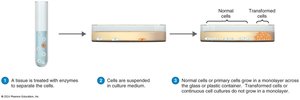
Cytopathic Effect
Viruses cause visible changes or deterioration in cell cultures, known as the cytopathic effect (CPE). This is a key method for viral identification.
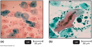
Viral Identification
Viruses are identified by:
Cytopathic effects in cell culture
Serological tests: ELISA detects viral antigens via antibody reactions
Nucleic acid tests: PCR amplifies viral genetic material
Viral Multiplication
Multiplication of Bacteriophages
Bacteriophages multiply via two main mechanisms:
Lytic cycle: Phage causes lysis and death of the host cell.
Lysogenic cycle: Phage DNA incorporates into host DNA, remains latent as a prophage.
Lytic Cycle Steps
Attachment: Phage attaches to host cell.
Penetration: Phage injects DNA into host.
Biosynthesis: Phage DNA and proteins produced.
Maturation: Assembly of new phage particles.
Release: Host cell lyses, releasing new phages.
Lysogenic Cycle Outcomes
Immunity to reinfection by the same phage
Phage conversion: Host cell acquires new properties (e.g., toxin production)
Specialized transduction: Specific bacterial genes transferred via phage
Multiplication of Animal Viruses
Animal viruses multiply through a series of steps:
Attachment: Virus binds to cell membrane.
Entry: Via receptor-mediated endocytosis or fusion.
Uncoating: Viral nucleic acid separated from capsid.
Biosynthesis: Production of viral nucleic acid and proteins.
Maturation: Assembly of viral components.
Release: By budding (enveloped viruses) or rupture (nonenveloped viruses).
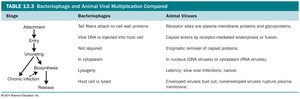
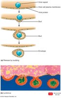
Biosynthesis of DNA and RNA Viruses
Biosynthesis of DNA Viruses
DNA viruses replicate their DNA in the nucleus of the host cell using host enzymes, while capsid proteins are synthesized in the cytoplasm.
Adenoviridae: Double-stranded DNA, nonenveloped; causes respiratory infections.
Poxviridae: Double-stranded DNA, enveloped; causes skin lesions (e.g., smallpox).
Herpesviridae: Double-stranded DNA, enveloped; includes viruses causing cold sores, chickenpox, mononucleosis, and Kaposi’s sarcoma.
Papovaviricetes: Double-stranded DNA, nonenveloped; causes warts and some cancers.
Hepadnaviridae: Double-stranded DNA, enveloped; hepatitis B virus, uses reverse transcriptase.
Biosynthesis of RNA Viruses
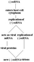
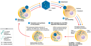
RNA viruses replicate in the cytoplasm using RNA-dependent RNA polymerase. The replication pathway depends on the type of RNA:
ssRNA (+) strand: Acts as mRNA for protein synthesis.
ssRNA (-) strand: Transcribed to a + strand to serve as mRNA.
dsRNA: Double-stranded RNA viruses.
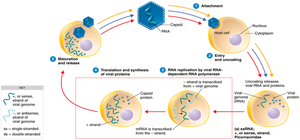
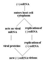
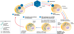
Examples of RNA Virus Families
Coronaviridae: Single-stranded RNA, + strand, enveloped; includes SARS-CoV-2.
Togaviridae: Single-stranded RNA, + strand, enveloped; includes alphaviruses.
Rhabdoviridae: Single-stranded RNA, - strand; includes rabies virus.
Picornaviridae: Single-stranded RNA, + strand, nonenveloped; includes enteroviruses, rhinovirus, hepatitis A.
Reoviridae: Double-stranded RNA, nonenveloped; includes rotavirus.
Biosynthesis of RNA Viruses That Use DNA
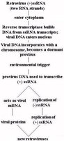
Retroviruses use reverse transcriptase to produce DNA from their RNA genome. The viral DNA integrates into the host chromosome as a provirus.
Retroviridae: Includes HIV and oncoviruses.
Viruses and Cancer
Oncogenic Viruses and Cell Transformation
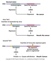
Some viruses can cause cancer by transforming normal cells into tumor cells. Proto-oncogenes are genes that regulate normal cell growth; when mutated, they become oncogenes and can induce cancer. Oncogenic viruses integrate into host DNA and induce tumors.
Sarcoma: Cancer of connective tissue.
Adenocarcinoma: Cancer of glandular epithelial tissue.
DNA Oncogenic Viruses
Adenoviridae
Herpesviridae (e.g., Epstein-Barr virus: Burkitt’s lymphoma)
Poxviridae
Papovaviridae (e.g., HPV: cervical and anal cancer)
Hepadnaviridae (e.g., hepatitis B virus)
RNA Oncogenic Viruses
Retroviridae: HTLV-1, HTLV-2 (adult T cell leukemia and lymphoma), FeLV (feline leukemia virus)
Oncolytic Viruses
Oncolytic viruses are used in cancer therapy to infect and kill tumor cells or stimulate an immune response against them.
Latent and Persistent Viral Infections
Latent Viral Infections
Latent viruses remain dormant in host cells for long periods and may reactivate due to changes in immunity. All herpesviruses are capable of latency.
Examples: HHV-1 (cold sores), Varicellovirus (shingles)
Persistent Viral Infections
Persistent infections progress slowly over time and are often fatal (e.g., subacute sclerosing panencephalitis caused by measles virus).
Plant Viruses and Viroids
Plant Viruses
Plant viruses enter through wounds or via insects, causing diseases in economically important plants. Plant cells are protected by an impermeable cell wall.
Viroids and Virusoids
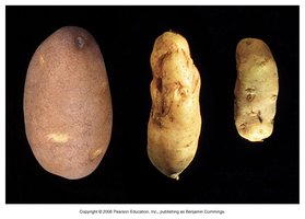
Viroids are short pieces of naked RNA that cause plant diseases, such as potato spindle tuber disease. Virusoids are viroids enclosed in a protein coat and require coinfection with a virus to cause disease.
Prions
Prion Diseases
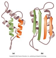
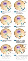
Prions are infectious proteins that cause neurodegenerative diseases. They are inherited or transmissible by ingestion, transplant, or surgical instruments. Prion diseases include spongiform encephalopathies such as "mad cow disease," Creutzfeldt-Jakob disease, and chronic wasting disease in deer.
Mechanism of Prion Infection
Prion diseases are caused by the conversion of normal cellular prion protein into a misfolded, infectious form that accumulates in brain cells, forming plaques.