 Back
BackViruses, Viroids, and Prions: Structure, Classification, and Replication
Study Guide - Smart Notes
 Tailored notes based on your materials, expanded with key definitions, examples, and context.
Viruses, Viroids, and Prions
General Characteristics of Viruses
Viruses are unique infectious agents that require a host cell for replication. They are fundamentally different from cellular life forms due to their simple structure and dependence on host machinery.
Obligatory intracellular parasites: Viruses must invade and take over a host cell to replicate.
Genetic material: Viruses contain either DNA or RNA, but never both.
Lack of cellular machinery: They do not possess ribosomes or ATP-generating mechanisms.
Protein coat: All viruses have a protein coat (capsid) that protects their genetic material.
Envelope and spikes: Some viruses are surrounded by an envelope derived from the host cell membrane and may have spikes for attachment and identification.
Host specificity: Most viruses infect only specific cell types in one host, determined by attachment sites and cellular factors.
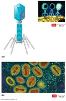
Bacteriophages: Viruses that infect bacteria are called bacteriophages.
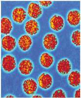
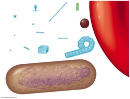
Size and Morphology of Viruses
Viruses are generally much smaller than bacteria and can be visualized only with electron microscopy. Their size and shape vary widely, influencing their classification and pathogenicity.
Size range: Viruses range from about 20 nm to 970 nm, much smaller than most bacteria.
Comparison: The largest viruses (e.g., poxviruses) are similar in size to the smallest bacteria, while the smallest viruses are much smaller than even a ribosome.
Viral Structure
The basic structural unit of a virus is the virion, which consists of nucleic acid surrounded by a protein coat. Some viruses also possess an envelope and surface projections called spikes.
Nucleic acid: Can be DNA or RNA, single- or double-stranded, linear or circular.
Capsid: Protein shell composed of subunits called capsomeres; determines the virus's shape and protects the genome.
Envelope: Lipid, protein, and carbohydrate layer derived from the host cell membrane; present in some animal viruses.
Spikes: Glycoprotein projections that aid in attachment and identification.
Viral Morphology
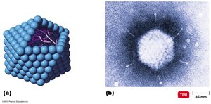
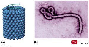
Viruses are classified by their morphology, which is determined by the arrangement of their capsid and presence or absence of an envelope.
Helical viruses: Rod-shaped, with nucleic acid coiled inside a helical capsid.
Polyhedral (Icosahedral) viruses: Capsid with 20 triangular faces, e.g., adenovirus.
Complex viruses: More intricate structures, such as bacteriophages with polyhedral heads and helical tails, or poxviruses with multiple coats.
Enveloped viruses: Capsid surrounded by an envelope, often with spikes.

Taxonomy and Classification of Viruses
Taxonomy of Viruses
Viruses are classified based on their genetic material, structure, and host range. The taxonomy follows a hierarchical system:
Family names: End in -viridae (e.g., Herpesviridae).
Genus names: End in -virus (e.g., Herpesvirus).
Species: Group of viruses sharing genetic information and ecological niche.
Subspecies: Designated by numbers (e.g., HHV-1, HHV-2).
Classification is based on properties such as nucleic acid type, symmetry, envelope presence, number of capsomeres, disease caused, and host.
Isolation, Cultivation, and Identification of Viruses
Growing Viruses
Viruses require living cells for growth and replication. Different methods are used for cultivating viruses depending on their host range.
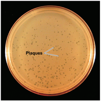
Bacteriophages: Grown on bacterial lawns, forming clear zones called plaques.
Animal viruses: Grown in living animals, embryonated eggs, or cell cultures (primary, diploid, or continuous cell lines).
Virus Identification
Viruses are identified using several laboratory techniques:
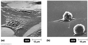
Cytopathic effects: Observable changes in host cells due to viral infection.
Serological tests: Detection of viral antigens or antibodies (e.g., neutralization tests, Western blot).
Nucleic acid analysis: Techniques such as PCR and RFLP for genetic identification.
Viral Multiplication
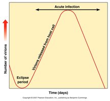
One-Step Growth Curve
The one-step growth curve illustrates the replication cycle of viruses, showing the eclipse period, rise period, and release of new virions.
Eclipse period: Time during which no infectious particles are detected as the virus is replicating inside the host cell.
Acute infection: Rapid increase in the number of virions as host cells are lysed and new viruses are released.
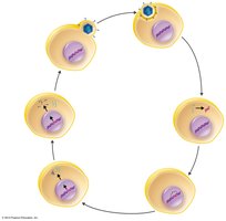
Lytic and Lysogenic Cycles in Bacteriophages
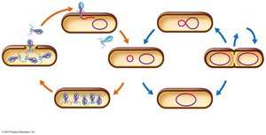
Bacteriophages can replicate via two main cycles: the lytic cycle, which destroys the host cell, and the lysogenic cycle, which integrates viral DNA into the host genome.
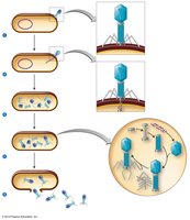
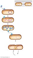
Lytic cycle: Involves attachment, penetration, biosynthesis, maturation, and release, resulting in host cell lysis.
Lysogenic cycle: Viral DNA integrates into the host chromosome as a prophage, replicating with the host cell and potentially conferring new traits (phage conversion).
Transduction
Transduction is a process by which bacteriophages transfer genetic material between bacteria, increasing genetic diversity.
Generalized transduction: Any bacterial gene may be transferred by a phage.
Specialized transduction: Only specific genes adjacent to the prophage are transferred.
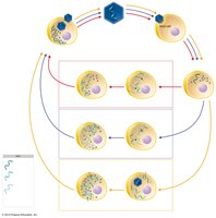
Animal Virus Multiplication
Steps in Animal Virus Multiplication
Animal viruses follow a similar replication process to bacteriophages, with some differences in entry and uncoating mechanisms.
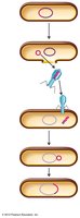
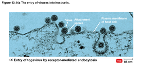
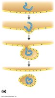
Attachment: Viruses attach to host cell membrane via spikes or fibers.
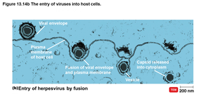
Penetration: Entry by endocytosis or fusion of the viral envelope with the host membrane.
Uncoating: Viral or host enzymes remove the capsid, releasing the viral genome.
Biosynthesis: Production of viral nucleic acids and proteins.
Maturation: Assembly of viral components into new virions.
Release: Virions exit the cell by budding (enveloped viruses) or rupture (nonenveloped viruses).
Biosynthesis of DNA and RNA Viruses
The replication strategies of DNA and RNA viruses differ based on their genome type.
DNA viruses: Replicate DNA in the nucleus and synthesize proteins in the cytoplasm. Assembly often occurs in the nucleus.
RNA viruses: Replicate and synthesize proteins in the cytoplasm. Positive-sense RNA can act as mRNA, while negative-sense RNA must be transcribed to mRNA first.
Retroviruses
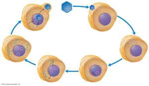
Retroviruses are RNA viruses that use reverse transcriptase to synthesize DNA from their RNA genome, which then integrates into the host genome as a provirus.
Reverse transcriptase: Enzyme that synthesizes DNA from RNA.
Integration: Viral DNA integrates into host DNA, where it can remain latent or direct synthesis of new viruses.
Viruses and Cancer
Oncogenes and Oncogenic Viruses
Some viruses can induce cancer by integrating their genetic material into the host genome, activating oncogenes that lead to uncontrolled cell growth.
Oncogenes: Genes that can cause a normal cell to become cancerous when activated.
Oncogenic viruses: Viruses that can activate oncogenes, including certain DNA and RNA viruses (e.g., Herpesviridae, Retroviridae).
Transformation: Process by which a normal cell becomes a cancer cell.
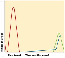
Latent and Persistent Viral Infections
Latent Infections
Latent infections occur when viruses remain dormant within host cells and can reactivate under certain conditions (e.g., herpes simplex virus causing cold sores).
Persistent Infections
Persistent infections involve continuous, slow production of virus over a long period, often leading to chronic disease (e.g., subacute sclerosing panencephalitis from measles virus).
Prions, Viroids, and Plant Viruses
Prions
Prions are infectious proteins that lack nucleic acids. They cause neurodegenerative diseases known as spongiform encephalopathies (e.g., Creutzfeldt-Jakob disease, mad cow disease).
Transmission: Inherited, or acquired by ingestion, transplant, or surgical instruments.
Viroids
Viroids are small, circular RNA molecules that infect plants, causing diseases such as potato spindle tuber disease. They lack a protein coat.
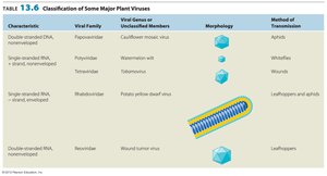
Plant Viruses
Plant viruses typically enter through wounds or are transmitted by insects. They can have diverse genome types and morphologies.
Characteristic | Viral Family | Viral Genus or Unclassified Members | Morphology | Method of Transmission |
|---|---|---|---|---|
Double-stranded DNA, nonenveloped | Papovaviridae | Cauliflower mosaic virus | Icosahedral | Aphids |
Single-stranded DNA, nonenveloped | Geminiviridae | Tomato yellow leaf curl virus | Icosahedral | Whiteflies |
Single-stranded RNA, nonenveloped | Potyviridae | Potato virus Y | Helical | Aphids |
Double-stranded RNA, nonenveloped | Reoviridae | Wound tumor virus | Icosahedral | Leafhoppers |
Summary Table: Major Virus Families Affecting Humans
Family | Genome Type | Envelope | Representative Diseases |
|---|---|---|---|
Parvoviridae | ssDNA | No | Fifth disease, anemia |
Adenoviridae | dsDNA | No | Respiratory infections, tumors in animals |
Papovaviridae | dsDNA | No | Warts, tumors, cancer |
Poxviridae | dsDNA | Yes | Smallpox, cowpox |
Herpesviridae | dsDNA | Yes | Herpes simplex, chickenpox, cytomegalovirus, Kaposi's sarcoma |
Hepadnaviridae | dsDNA | Yes | Hepatitis B |
Picornaviridae | ssRNA (+) | No | Polio, coxsackievirus, rhinovirus, hepatitis A |
Caliciviridae | ssRNA (+) | No | Norovirus, hepatitis E |
Togaviridae | ssRNA (+) | Yes | Rubella, alphaviruses |
Flaviviridae | ssRNA (+) | Yes | Yellow fever, dengue, hepatitis C |
Coronaviridae | ssRNA (+) | Yes | Common cold, SARS |
Rhabdoviridae | ssRNA (−) | Yes | Rabies |
Filoviridae | ssRNA (−) | Yes | Ebola, Marburg |
Paramyxoviridae | ssRNA (−) | Yes | Mumps, measles, parainfluenza |
Deltaviridae | ssRNA (−) | No | Hepatitis D (requires coinfection with Hepadnavirus) |
Orthomyxoviridae | ssRNA (−), segmented | Yes | Influenza |
Bunyaviridae | ssRNA (−), segmented | Yes | Hantavirus, CE virus |
Arenaviridae | ssRNA (−), segmented | Yes | Lassa fever, VEE |
Retroviridae | ssRNA (+), diploid | Yes | HIV, RNA tumor viruses |
Reoviridae | dsRNA | No | Rotavirus, Colorado tick fever |
