 Back
Viruses, Viroids, and Prions: Structure, Replication, and Infection
Study Guide - Smart Notes
 Tailored notes based on your materials, expanded with key definitions, examples, and context.
Viruses, Viroids, and Prions
Introduction to Viruses
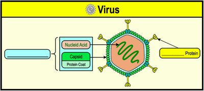
Viruses are obligate intracellular parasites composed of either DNA or RNA, surrounded by a protein coat, and sometimes a lipid envelope. They require a host cell to replicate and are responsible for a wide range of diseases in animals, plants, and bacteria.
Capsid: The protein coat that protects viral nucleic acids from environmental toxins and enzymes.
Nucleocapsid: Combination of nucleic acids and the capsid.
Spikes: Surface proteins that allow viruses to bind to specific host cell receptors to initiate infection.
Virion: A complete virus particle released from the host cell into the extracellular space.
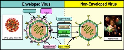
Enveloped vs. Non-Enveloped Viruses
Viruses can be classified based on the presence or absence of a lipid envelope.
Enveloped Viruses: Have an outer lipid bilayer obtained from the host cell. A matrix protein links the envelope to the nucleocapsid. These viruses are susceptible to soaps and detergents, which damage the envelope and render the virus non-infectious. Example: SARS-CoV-2 (COVID-19).
Non-Enveloped (Naked) Viruses: Lack an outer lipid bilayer and are generally more resistant to environmental stress.
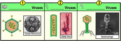
Shapes of Viruses
Viruses exhibit several common shapes, which are important for their classification and infection mechanisms.
Icosahedral: Spherical appearance with 20 triangular sides (e.g., Human Rhinovirus).
Helical: Cylindrical shape with capsids arranged in a spiral (e.g., Ebola Virus).
Complex: Multiple shapes, often seen in bacteriophages.


Plaque Assays
Determining Viral Concentration
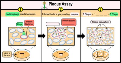
Plaque assays are used to quantify the number of phage particles in a sample by observing the formation of plaques on a layer of cultured cells.
Plaques: Clear areas in a layer of cultured cells indicating cell lysis from phage infections.
Plaque-Forming Unit (PFU): Each plaque is presumed to arise from a single lytic virus particle.
Titer: The concentration of phage particles in the original undiluted sample.
Animal Virus Infections
Steps of Animal Virus Infection
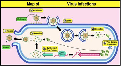
Animal virus infection follows a series of steps, each critical for successful viral replication.
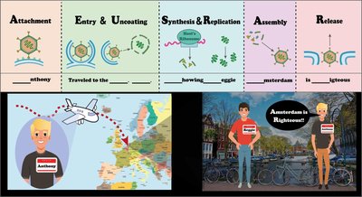
Attachment: Virus binds to host cell receptors via spike proteins.
Entry: Virus enters the host cell.
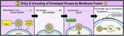
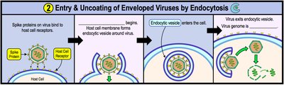
Uncoating: Viral genome is released from the protein coat into the host cell cytoplasm.
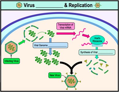
Synthesis & Replication: Host cell machinery synthesizes viral proteins and replicates the viral genome.
Assembly: New viral components are assembled into virions.
Release: New virions are released from the host cell.
Bacteriophage Infections
Lytic Cycle
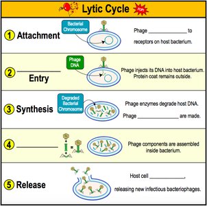
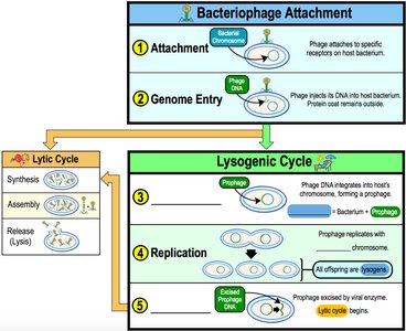
The lytic cycle is a productive infection that results in the destruction of the host cell and the release of new phages.
Attachment: Phage attaches to specific receptors on the host bacterium.
Entry: Phage injects its DNA into the host bacterium.
Synthesis: Phage enzymes degrade host DNA and viral components are synthesized.
Assembly: New phage particles are assembled inside the bacterium.
Release: Host cell lyses, releasing new infectious bacteriophages.
Lysogenic Cycle
Temperate phages may not immediately produce new phages. Instead, their DNA integrates into the host genome, forming a prophage.
Attachment & Genome Entry: Same as lytic cycle.
Integration: Phage DNA integrates into the host chromosome.
Replication: Prophage replicates with the host genome.
Excision: Prophage may exit the host genome and enter the lytic cycle.
Lysogenic Conversion
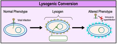
Lysogenic conversion refers to the phenotypic change of a lysogen based on the prophage it carries.
Can result in altered surface structures and immunity to superinfection.
May confer toxin-producing properties to the host cell.
Animal Virus Attachment, Entry, and Uncoating
Attachment to Host Cell
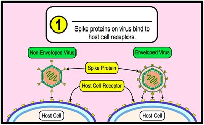
Animal virus attachment is similar to bacteriophage attachment. Spike proteins on the virus recognize and bind to specific host cell receptors, allowing entry.
Binding to these receptors is essential for viral entry.
Entry & Uncoating in the Host Cell
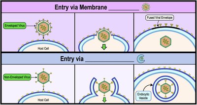
Enveloped viruses enter host cells via membrane fusion or endocytosis, while non-enveloped viruses enter only via endocytosis.
Membrane Fusion: Lipid envelope fuses with host cell membrane.
Endocytosis: Host cell forms an endocytic vesicle around the virus.
Animal Virus Synthesis & Replication
Overview of Synthesis & Replication
After entry and uncoating, viral synthesis and replication begin. The mechanism depends on the type of viral genome.
Structural proteins and replication enzymes are synthesized.
Viral genome is replicated multiple times inside the host cell.
Types of Animal Virus Genomes
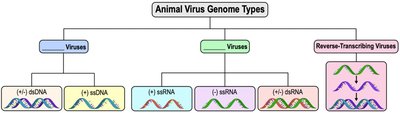
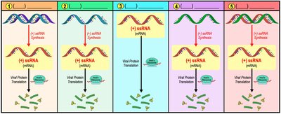
The synthesis and replication mechanism depends on the viral genome type:
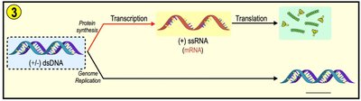
(+/–) dsDNA: Double-stranded DNA, follows central dogma.
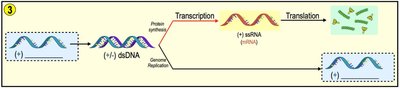
(+) or (–) ssDNA: Single-stranded DNA converted to dsDNA before replication.
(+) ssRNA: Coding single-stranded RNA, directly translated by host ribosomes.
(–) ssRNA: Non-coding single-stranded RNA, used as template to synthesize (+) ssRNA.
(+/–) dsRNA: Double-stranded RNA, template for (+) ssRNA.
DNA Virus Synthesis & Replication
Most DNA viruses replicate inside the host cell nucleus using host cell components. They encode their own DNA polymerases for genome replication.
dsDNA virus replication follows the central dogma: DNA → mRNA → Protein.
ssDNA viruses must first convert ssDNA to dsDNA before transcription.
RNA Virus Synthesis & Replication
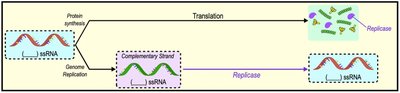
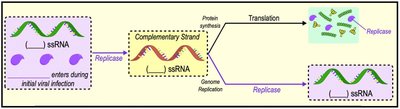
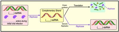
Most RNA viruses replicate in the cytoplasm and require a viral RNA polymerase (replicase).
(+) ssRNA is directly translated by host ribosomes.
(–) ssRNA must bring replicase enzyme during infection to synthesize (+) ssRNA.
dsRNA viruses use replicase to make (+) ssRNA for translation.
Acute vs. Persistent Viral Infections
Categories of Animal Virus Infections
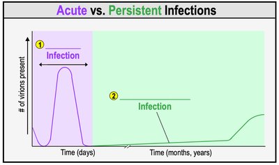
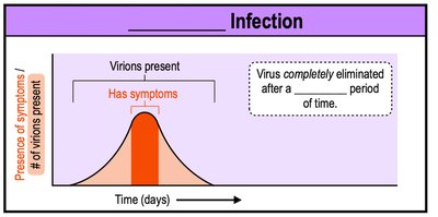
Animal virus infections are categorized as acute or persistent.
Acute Infections: Sudden onset of symptoms, short duration, virus eliminated by immune system.
Persistent Infections: Last for years or the entire life of the host. Includes chronic and latent infections.
COVID-19 Virus (SARS-CoV-2)
Characteristics and Infection
COVID-19 is an enveloped, (+) ssRNA virus with spike proteins giving it a crown-like appearance. It causes acute infections, primarily affecting lung tissue and is transmitted via respiratory droplets and poor hygiene.
Enveloped viruses are easier to deactivate with soaps and disinfectants.
SARS-CoV-2 genome can be directly translated by host ribosomes.
Plant Viruses
Structure and Transmission
Plant viruses are similar to animal viruses in morphology and nucleic acid types, but most are non-enveloped. They enter host cells via wound sites and are transmitted by insects, soil, contaminated seeds, or humans.
Infection of adjacent plant cells occurs via plasmodesmata.
Viroids
Structure and Infection
Viroids are obligate intracellular parasites made of a single, short strand of ssRNA forming a closed ring. They infect only plant cells, entering via wound sites and replicating using host RNA-dependent RNA polymerase.
Viroids do not encode proteins but act as small interfering RNAs, blocking plant gene expression.
Prions
Structure and Disease
Prions are proteinaceous infectious agents composed of misfolded proteins. They cause normal proteins to misfold, leading to neurodegenerative diseases such as transmissible spongiform encephalopathies (e.g., Scrapie, Mad Cow Disease).
Prions accumulate in neural tissue, causing brain tissue to deteriorate.
Prion diseases are difficult to deactivate and have no cure.
