 Back
BackViruses, Viroids, and Prions: Structure, Replication, and Disease
Study Guide - Smart Notes
 Tailored notes based on your materials, expanded with key definitions, examples, and context.
Viruses: Structure and Classification
Comparing Cells to Viruses
Viruses are fundamentally different from cellular life forms. They are acellular, obligate intracellular parasites that require a host cell for replication. Unlike cells, viruses do not metabolize, grow, or divide independently.
Viruses: Acellular, contain either DNA or RNA, replicate using host machinery, have a protein capsid, and may possess an envelope.
Cells: Cellular, contain both DNA and RNA, self-replicate, metabolize, and grow.
Obligate intracellular parasites: Viruses must infect a host cell to reproduce.
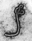
Relative Sizes of Viruses
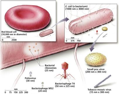
Viruses are ultramicroscopic, typically ranging from 10 nm to over 500 nm, much smaller than most cells and organelles.
Examples: Poliovirus (30 nm), Smallpox virus (200 nm x 300 nm), Tobacco mosaic virus (15 nm x 300 nm).
Cells: E. coli (1000 nm x 3000 nm), Red blood cell (10,000 nm diameter).
Structure of Viruses
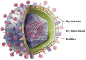
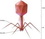
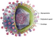
Viruses exist in two states: extracellular (virion) and intracellular. The virion consists of a nucleic acid genome surrounded by a protein capsid, and some have an envelope derived from the host cell.
Capsid: Protein shell protecting the genome.
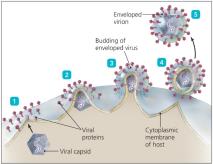
Envelope: Phospholipid bilayer with viral proteins, acquired from the host cell during replication.
Glycoprotein spikes: Facilitate attachment to host cells.
The Viral Envelope
Some animal viruses possess an envelope, which is acquired from the host cell during viral replication. The envelope contains viral glycoproteins essential for host cell recognition and attachment.
Enveloped viruses are more fragile than non-enveloped viruses.
Envelope proteins play a key role in immune evasion and host specificity.
Hosts of Viruses
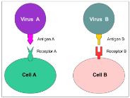
Viruses exhibit host specificity, determined by the affinity of viral surface proteins for complementary host cell receptors.
Most viruses infect a particular type of cell or host (narrow host range).
Some are generalists, infecting multiple cell types or hosts (rare).
Genetic Material of Viruses
Viruses display remarkable diversity in their genomes, which may be DNA or RNA, single- or double-stranded, linear, circular, or segmented.
Genome types: dsDNA, ssDNA, dsRNA, ssRNA (+ or −).
Genome size is much smaller than cellular genomes.

Other Ways to Categorize Viruses
Viruses are classified based on nucleic acid structure, host range, size, shape, capsid structure, and presence or absence of an envelope.
Capsid shapes: Helical, polyhedral, complex.
Capsomeres: Protein subunits of the capsid.
Viral Replication Mechanisms
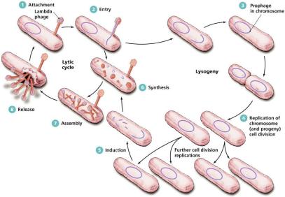
Lytic Replication
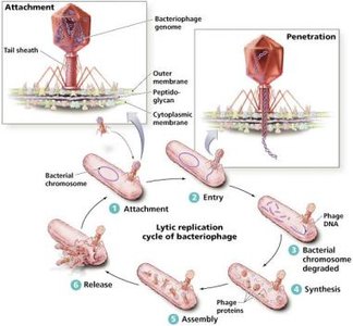
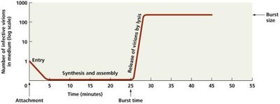
Lytic replication is a destructive process resulting in the death and lysis of the host cell. It consists of five steps: attachment, entry, synthesis, assembly, and release.
Burst time: Time required to complete the lytic cycle.
Burst size: Number of new virions released.
Lysogenic and Latent Replication
Lysogenic replication involves integration of the viral genome into the host chromosome, resulting in a period of dormancy. Latent replication occurs in animal viruses, allowing them to remain dormant as proviruses.
Provirus incorporation is permanent in animal cells.
Viruses can evade the immune system during latency.
Replication of Animal Viruses
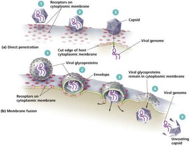
Animal viruses may enter cells by direct penetration, membrane fusion, or phagocytosis. The replication strategy depends on the type of nucleic acid.
DNA viruses often replicate in the nucleus.
RNA viruses typically replicate in the cytoplasm.
Synthesis of Animal Viruses
The synthesis of viral components varies by genome type. Positive-sense RNA can act as mRNA, while negative-sense RNA requires synthesis of a complementary strand.
Retroviruses use reverse transcriptase to synthesize DNA from RNA.
DNA viruses: dsDNA replication is similar to cellular DNA replication.
ssDNA viruses: Parvoviruses fold ssDNA to form dsDNA for replication.
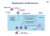
Assembly and Release of Animal Viruses
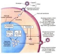
Most DNA viruses assemble in the nucleus, while RNA viruses assemble in the cytoplasm. Enveloped viruses are released by budding, causing persistent infections; non-enveloped viruses are released by lysis or exocytosis.
Number of viruses produced depends on virus type and host cell health.
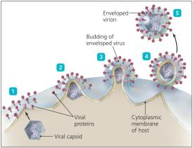
Bacteriophage vs. Animal Virus Replication
Step | Bacteriophage | Animal Virus |
|---|---|---|
Attachment | Proteins on tails attach to cell wall | Spikes, capsids, or envelope proteins attach to cell membrane |
Penetration | Genome injected or diffuses into cell | Capsid enters by penetration, fusion, or endocytosis |
Uncoating | None | Capsid removed by cell enzymes |
Site of Synthesis | Cytoplasm | RNA viruses: cytoplasm; DNA viruses: nucleus |
Site of Assembly | Cytoplasm | RNA viruses: cytoplasm; DNA viruses: nucleus |
Release | Lysis | Naked virions: exocytosis or lysis; enveloped virions: budding |
Chronic Infection | Lysogeny, always incorporated into host chromosome | Latency, with or without incorporation into host DNA |
Example Viruses
Human Immunodeficiency Virus (HIV)
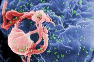
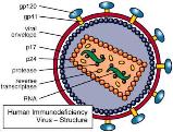
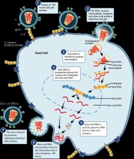
HIV is a retrovirus with a double-stranded RNA genome and three unique enzymes: reverse transcriptase, integrase, and protease. It infects CD4 T-cells and macrophages.
Attachment: gp120 binds to CD4.
Entry: Penetration and reverse transcription.
Integration: Viral DNA integrates into host chromosome.
Synthesis: Biosynthesis of viral RNA and proteins.
Assembly: New virus assembled with protease.
Release: Virions released from host cell.
Influenza Virus
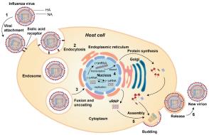
Influenza virus carries RNA-dependent RNA polymerase and has a genome of 8 ssRNA molecules. Glycoproteins hemagglutinin (H) and neuraminidase (N) are key for attachment and release.
Attachment: H binds to sialic acid on host cell.
Entry: Endocytosis and penetration of vRNA into nucleus.
Synthesis: Biosynthesis of vRNA and proteins.
Assembly: New virus assembled.
Release: N cleaves sialic acid for viral exit.
Antigenic drift: Minor changes in H and N.
Antigenic shift: Major changes via gene reassortment, leading to pandemics.
Viruses and Cancer
The Role of Viruses in Cancer
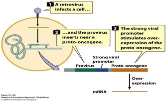
Viruses can cause cancer by disrupting normal cell cycle regulation. DNA and RNA viruses are implicated in ~25% of human cancers.
Examples: Epstein-Barr virus (Burkitt’s lymphoma), HPV (cervical cancer), HBV/HCV (liver cancer), HIV (Kaposi sarcoma), HHV-8 (Kaposi sarcoma), HTLV-1 (adult T-cell leukemia/lymphoma).
Mechanisms: Insertion into tumor suppressor genes, increased transcription of oncogenes, carrying oncogenes in viral genome.

Other Parasitic Particles: Viroids and Prions
Viroids
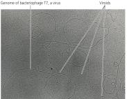
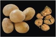
Viroids are small, circular ssRNA molecules that infect plants. They lack a capsid and do not code for proteins, but cause disease by interfering with plant RNA.
Viroid RNA adheres to complementary plant RNA, leading to degradation and disease.
Prions
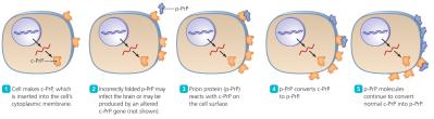
Prions are infectious proteins capable of inducing abnormal folding of normal cellular PrP proteins, leading to fatal neurological diseases.
Normal PrP: α-helices, involved in copper transport in neurons.
Prion PrP: β-pleated sheets, disease-causing.
Transmission: Ingestion, transplantation, or contact with infected tissues.
Diseases: BSE (cows), Scrapie (sheep), CWD (deer/elk), Kuru, vCJD (humans).
No standard treatment; prions are destroyed only by incineration.
Comparison Table: Bacteria, Viruses, Viroids, Prions
Property | Bacteria | Viruses | Viroids | Prions |
|---|---|---|---|---|
Width | 200–2000 nm | 10–400 nm | 2 nm | 5 nm |
Length | 200–550,000 nm | 20–800 nm | 40–130 nm | 5 nm |
Nucleic Acid | DNA & RNA | DNA or RNA | RNA only | None |
Protein | Present | Present | Absent | Present (PrP) |
Cellular | Yes | No | No | No |
Cytoplasmic Membrane | Present | Absent (some viruses have envelope) | Absent | Absent |
Functional Ribosomes | Present | Absent | Absent | Absent |
Growth | Present | Absent | Absent | Absent |
Self-Replicating | Yes | No | No | No; transforms PrP already present |
Responsiveness | Present | Some bacteriophages respond to host | Absent | Absent |
Metabolism | Present | Absent | Absent | Absent |
Families of Human Viruses
DNA Viruses
Family | Strand Type | Representative Genera (Diseases) |
|---|---|---|
Poxviridae | Double | Orthopoxvirus (smallpox) |
Herpesviridae | Double | Simplexvirus (herpes), Varicellovirus (chicken pox), Epstein-Barr virus (mono, lymphoma), Cytomegalovirus (birth defects), Roseolovirus (roseola) |
Papillomaviridae | Double | Papillomavirus (warts, cervical/penile cancers) |
Polyomaviridae | Double | Polyomavirus (leukoencephalopathy) |
Adenoviridae | Double | Mastadenovirus (conjunctivitis, respiratory infections) |
Hepadnaviridae | Partial single/double | Orthohepadnavirus (hepatitis B) |
Parvoviridae | Single | Erythrovirus (erythema infectiosum) |
RNA Viruses
Family | Strand Type | Representative Genera (Diseases) |
|---|---|---|
Picornaviridae | Single, + | Enterovirus (polio), Hepatovirus (hepatitis A) |
Caliciviridae | Single, + | Norovirus (gastroenteritis) |
Astroviridae | Single, + | Astrovirus (gastroenteritis) |
Hepeviridae | Single, + | Hepevirus (hepatitis E) |
Togaviridae | Single, + | Alphavirus (encephalitis), Rubivirus (rubella) |
Flaviviridae | Single, + | Flavivirus (yellow fever, encephalitis), Hepacivirus (hepatitis C) |
Coronaviridae | Single, + | Coronavirus (common cold, SARS) |
Retroviridae | Single, +, segmented | Deltaretrovirus (leukemia), Lentivirus (AIDS) |
Paramyxoviridae | Single, − | Paramyxovirus (cold), Pneumovirus (pneumonia), Morbillivirus (measles), Rubulavirus (mumps) |
Rhabdoviridae | Single, − | Lyssavirus (rabies) |
Filoviridae | Single, − | Filovirus (Ebola, Marburg) |
Bunyaviridae | Single, −, segmented | Bunyavirus (encephalitis), Hantavirus (pneumonia) |
Orthomyxoviridae | Single, −, segmented | Influenzavirus (flu) |
Arenaviridae | Single, −, segmented | Lassavirus (hemorrhagic fever) |
Reoviridae | Double, segmented | Orbivirus (encephalitis), Rotavirus (diarrhea), Coltivirus (tick fever) |
Additional info: Positive-sense (+RNA) is equivalent to mRNA; negative-sense (−RNA) is complementary to mRNA and cannot be directly translated.
