 Back
BackViruses, Viroids, and Prions: Structure, Replication, and Pathogenicity
Study Guide - Smart Notes
 Tailored notes based on your materials, expanded with key definitions, examples, and context.
Chapter 13: Viruses, Cancer, and Prions
Introduction to Viruses
Viruses are minuscule, acellular infectious agents that carry genetic material in the form of DNA or RNA. Unlike cells, viruses are obligate intracellular parasites, meaning they require a host cell to replicate and cannot carry out metabolic processes independently.
Acellular: Viruses lack cellular structure and do not possess cytoplasmic membranes, cytosol, or organelles.
Obligate Intracellular Parasites: They must infect a host cell to reproduce, utilizing the host's metabolic machinery.
Genome: Viral genomes may be DNA or RNA, single- or double-stranded, linear, circular, or segmented.
Size: Viruses are typically much smaller than cells, ranging from 10 nm to over 500 nm.
Comparing Cells and Viruses
Cells and viruses differ fundamentally in structure and function. The table below summarizes key differences:
Feature | Cells | Viruses |
|---|---|---|
Cellular Structure | Yes | No |
Metabolism | Present | Absent |
Growth | Present | Absent |
Reproduction | Self-replicating | Dependent on host |
Genetic Material | DNA and RNA | DNA or RNA (never both) |
Size | 200 nm – 12 cm | 10 nm – 500 nm |
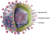
Structure of Viruses
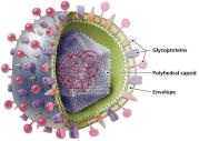
Viruses exist in two states: extracellular (virion) and intracellular. The virion consists of a nucleic acid core surrounded by a protein coat (capsid), and in some cases, an envelope derived from the host cell membrane.
Capsid: Protein shell that protects the viral genome and aids in attachment to host cells.
Envelope: Present in some animal viruses, composed of a phospholipid bilayer and viral proteins (often glycoprotein spikes).
Enzymes: Some viruses carry enzymes necessary for their replication cycle.
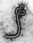
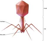
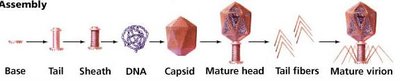
Viral Morphologies
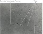
Viruses display a variety of shapes, including helical, polyhedral, and complex forms. Bacteriophages, viruses that infect bacteria, often have a complex structure with a head, tail, and tail fibers.
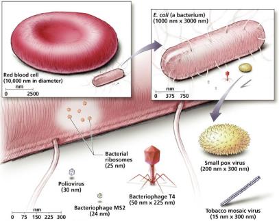
Relative Sizes of Viruses
Viruses are much smaller than most cells and organelles. The following image compares the sizes of various viruses, bacteria, and eukaryotic cells.
Viral Envelopes
Some animal viruses possess an envelope acquired from the host cell during replication. The envelope contains viral glycoproteins that facilitate host cell recognition and entry. Enveloped viruses are generally more sensitive to environmental conditions than nonenveloped viruses.
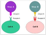
Host Range and Specificity
Viruses exhibit specificity for their host due to interactions between viral surface proteins and host cell receptors. Most viruses infect only specific cell types or species, though some are generalists.
Genetic Material of Viruses
Viral genomes are highly diverse and are a primary means of classification. They may be:
DNA or RNA (never both)
Single-stranded (ss) or double-stranded (ds)
Linear, circular, or segmented
Positive-sense (+) or negative-sense (−) RNA

Classification of Viruses
Viruses are classified based on:
Type of nucleic acid (DNA or RNA, ss or ds)
Host range
Size and shape
Capsid structure
Presence or absence of an envelope
Viral Replication
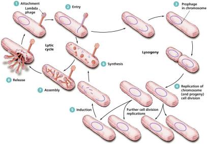
Lytic Replication Cycle
The lytic cycle is a replication process resulting in the destruction of the host cell. It consists of five steps:
Attachment: Virus binds to host cell surface.
Entry: Viral genome enters the host cell.
Synthesis: Host machinery synthesizes viral components.
Assembly: New virions are assembled.
Release: Host cell lyses, releasing new virions.
Lysogenic and Latent Replication
Some viruses can integrate their genome into the host's DNA, entering a dormant state (lysogeny in bacteriophages, latency in animal viruses). The viral genome, called a prophage or provirus, can reactivate later to enter the lytic cycle.
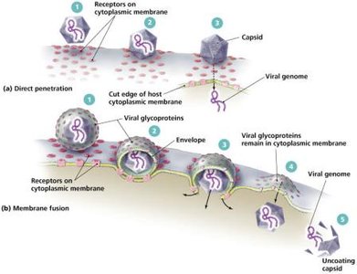
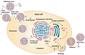
Replication of Animal Viruses
Animal viruses may enter host cells by direct penetration, membrane fusion, or phagocytosis. The replication strategy depends on the type of viral genome (DNA or RNA, ss or ds).
DNA viruses: Often replicate in the nucleus.
RNA viruses: Usually replicate in the cytoplasm.
Retroviruses: Use reverse transcriptase to synthesize DNA from RNA.
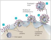
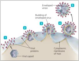
Assembly and Release of Animal Viruses
Most DNA viruses assemble in the nucleus, while RNA viruses assemble in the cytoplasm. Enveloped viruses are released by budding, causing persistent infections, while nonenveloped viruses are released by lysis or exocytosis.
Latent Replication of Animal Viruses
Latent viruses (proviruses) can remain dormant within host cells, sometimes for years, evading the immune system. If integrated into host DNA, the provirus becomes a permanent part of the host genome.
Bacteriophage vs. Animal Virus Replication
Step | Bacteriophage | Animal Virus |
|---|---|---|
Attachment | Proteins on tails attach to cell wall | Spikes, capsids, or envelope proteins attach to cell membrane |
Penetration | Genome injected or diffuses into cell | Capsid enters by penetration, fusion, or endocytosis |
Uncoating | None | Capsid removed by enzymes |
Site of Synthesis | Cytoplasm | RNA viruses: cytoplasm; DNA viruses: nucleus |
Release | Lysis | Exocytosis, lysis, or budding |
Chronic Infection | Lysogeny | Latency |
Examples of Viruses
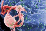
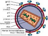
Human Immunodeficiency Virus (HIV)
HIV is a retrovirus with a double-stranded RNA genome. It carries reverse transcriptase, integrase, and protease enzymes, infecting CD4 T-cells and macrophages. The replication cycle involves attachment, entry, reverse transcription, integration, synthesis, assembly, and release.
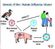
Influenza Virus
Influenza is an RNA virus with a segmented genome and envelope glycoproteins hemagglutinin (H) and neuraminidase (N). Antigenic drift (minor changes) and shift (major changes) in these proteins can lead to new strains and pandemics.
Viruses and Cancer
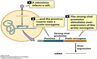
Oncogenic Viruses
Some viruses can cause cancer by disrupting normal cell cycle regulation. Mechanisms include insertional mutagenesis (disrupting tumor suppressor genes), upregulation of oncogenes, or carrying viral oncogenes.
Examples: Epstein-Barr virus, Human papillomavirus (HPV), Hepatitis B and C viruses, HIV, Human herpesvirus 8, HTLV-1.

Other Parasitic Particles: Viroids and Prions
Viroids
Viroids are small, circular, single-stranded RNA molecules that infect plants. They lack a protein coat and do not code for proteins, causing disease by interfering with plant RNA.

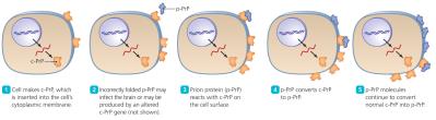
Prions
Prions are infectious proteins that cause neurodegenerative diseases. The normal cellular form (PrPC) has α-helices, while the disease-causing form (PrPSc) has β-pleated sheets. Prions propagate by inducing misfolding of normal proteins.
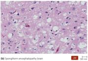
Diseases: Bovine spongiform encephalopathy (BSE), Scrapie, Chronic wasting disease, Kuru, Creutzfeldt-Jakob disease (CJD).
Treatment: Prions are resistant to standard sterilization; only incineration is effective.
Comparison Table: Bacteria, Viruses, Viroids, Prions
Feature | Bacteria | Viruses | Viroids | Prions |
|---|---|---|---|---|
Width | 200–2000 nm | 10–400 nm | 2 nm | 5 nm |
Length | 200–550,000 nm | 20–800 nm | 40–130 nm | 5 nm |
Nucleic Acid | DNA and RNA | DNA or RNA | RNA only | None |
Protein | Present | Present | Absent | Present (PrP) |
Cellular | Yes | No | No | No |
Cytoplasmic Membrane | Present | Absent (some have envelope) | Absent | Absent |
Functional Ribosomes | Present | Absent | Absent | Absent |
Growth | Present | Absent | Absent | Absent |
Self-Replicating | Yes | No | No | No; transforms PrP protein |
Responsiveness | Present | Some bacteriophages respond | Absent | Absent |
Metabolism | Present | Absent | Absent | Absent |
Families of Human Viruses
DNA Viruses
Family | Strand Type | Representative Genera (Diseases) |
|---|---|---|
Poxviridae | Double | Orthopoxvirus (smallpox) |
Herpesviridae | Double | Simplexvirus (herpes), Varicellovirus (chickenpox), Epstein-Barr virus (mononucleosis, Burkitt’s lymphoma) |
Papillomaviridae | Double | Papillomavirus (warts, cervical cancer) |
Polyomaviridae | Double | Polyomavirus (leukoencephalopathy) |
Adenoviridae | Double | Mastadenovirus (conjunctivitis, respiratory infections) |
Hepadnaviridae | Partial single/double | Orthohepadnavirus (hepatitis B) |
Parvoviridae | Single | Erythrovirus (erythema infectiosum) |
RNA Viruses
Family | Strand Type | Representative Genera (Diseases) |
|---|---|---|
Picornaviridae | Single, + | Enterovirus (polio), Hepatovirus (hepatitis A) |
Caliciviridae | Single, + | Norovirus (gastroenteritis) |
Astroviridae | Single, + | Astrovirus (gastroenteritis) |
Hepeviridae | Single, + | Hepevirus (hepatitis E) |
Togaviridae | Single, + | Alphavirus (encephalitis), Rubivirus (rubella) |
Flaviviridae | Single, + | Flavivirus (yellow fever), Hepacivirus (hepatitis C) |
Coronaviridae | Single, + | Coronavirus (common cold, SARS) |
Retroviridae | Single, +, segmented | Deltaretrovirus (leukemia), Lentivirus (AIDS) |
Paramyxoviridae | Single, − | Paramyxovirus (common cold), Morbillivirus (measles) |
Rhabdoviridae | Single, − | Lyssavirus (rabies) |
Filoviridae | Single, − | Filovirus (Ebola), Marburgvirus |
Bunyaviridae | Single, −, segmented | Bunyavirus, Hantavirus |
Orthomyxoviridae | Single, −, segmented | Influenzavirus (flu) |
Arenaviridae | Single, −, segmented | Lassavirus |
Reoviridae | Double, segmented | Orbivirus, Rotavirus |
