 Back
Visualization of Bacteria: Microscopy and Staining Techniques
Study Guide - Smart Notes
 Tailored notes based on your materials, expanded with key definitions, examples, and context.
Visualization of Bacteria
Units of Measurement in Microbiology
Microorganisms are measured using the metric system, primarily in micrometers (µm) and nanometers (nm). Understanding these units and their conversions is essential for interpreting microscopic observations.
Micrometer (µm): 1 µm = 10-6 meters = 10-3 millimeters
Nanometer (nm): 1 nm = 10-9 meters = 10-6 millimeters
1,000 nm = 1 µm
0.001 µm = 1 nm
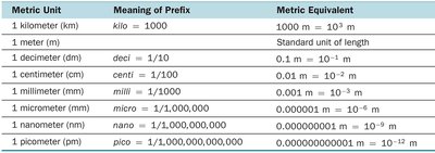
Example: 10 millimeters (mm) = 10,000,000 nanometers (nm).
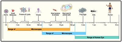
Cell Size and Surface Area-to-Volume Ratio
The size and shape of cells are closely related to their function. Small cells have a higher surface area-to-volume ratio, which allows for more efficient nutrient uptake and waste elimination, supporting faster growth and division.
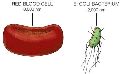
Average bacterial cell: 0.5–2.0 µm in diameter
Red blood cell: 7.5 µm in diameter
Large surface area relative to volume enables efficient transport of materials.
Surface Area-to-Volume Ratio: As cells increase in size, their volume grows faster than their surface area, reducing the efficiency of nutrient exchange.
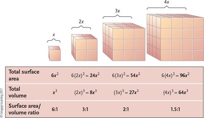
Example: Small cells (higher ratio) grow and divide faster than large cells (lower ratio).
Microscopy: Observing Microorganisms
History of Microscopy
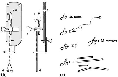
Anton van Leeuwenhoek (1674) was the first to observe microorganisms using a simple microscope with a single lens. Modern microscopes use multiple lenses for greater magnification and resolution.
Compound Light Microscope
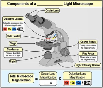
A compound microscope uses two or more lenses to magnify specimens. The total magnification is the product of the magnifications of the objective and ocular lenses.
Total Magnification:
Resolution: The minimum distance at which two points can be distinguished as separate.
Contrast: The difference in light intensity between the specimen and the background.
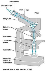
Key Point: Shorter wavelengths of light provide greater resolution.
Parts of a Light Microscope
Understanding the components of a light microscope is essential for proper use and maintenance.
Ocular lens (eyepiece): Magnifies the image further
Objective lenses: Provide primary magnification (e.g., 4x, 10x, 40x, 100x)
Stage: Holds the specimen slide
Condenser: Focuses light onto the specimen
Illuminator: Light source
Types of Light Microscopes
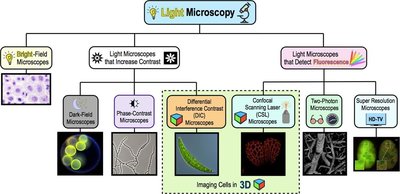
Several types of light microscopes are used to visualize microorganisms, each with unique advantages for contrast and detail.
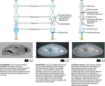
Bright-field microscope: Produces a dark image against a bright background; best for stained specimens.
Dark-field microscope: Enhances contrast in unstained samples; specimen appears bright against a dark background.
Phase-contrast microscope: Increases contrast in transparent specimens without staining.
Fluorescence microscope: Uses fluorescent dyes to visualize specific structures.
Electron Microscopy
Electron microscopes use beams of electrons instead of light, allowing for much higher resolution and magnification. There are two main types:
Transmission Electron Microscope (TEM): Electrons pass through thin sections of a specimen, revealing internal structures in 2D images.
Scanning Electron Microscope (SEM): Electrons are reflected from the surface, producing detailed 3D images of external structures.
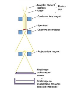
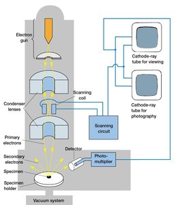
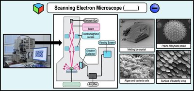
Applications: TEM is used for studying internal cell structures, while SEM is ideal for examining surface details.
Staining Techniques in Microbiology
Preparation and Fixation of Specimens
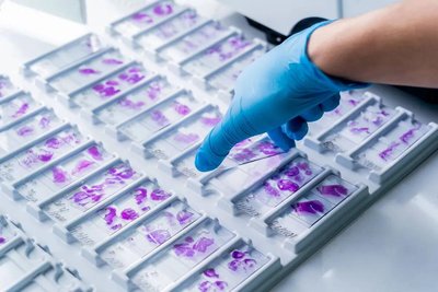
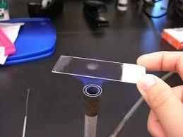
Staining increases the visibility of microorganisms and highlights specific structures. Fixation preserves cell morphology and attaches cells to the slide.
Heat fixation: Preserves overall morphology but not internal structures.
Chemical fixation: Preserves fine cellular substructure, suitable for delicate organisms.
Simple Staining
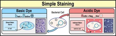
Simple staining uses a single dye to highlight the entire microorganism, making it easier to observe cell shape and arrangement.
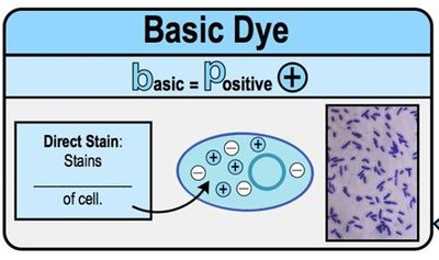
Basic dyes: Positively charged; stain the cell directly (e.g., crystal violet).
Acidic dyes: Negatively charged; stain the background (negative staining).
Differential Staining
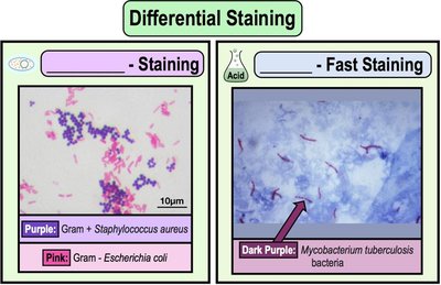
Differential stains distinguish between different groups of bacteria based on their staining properties. The most common are the Gram stain and the acid-fast stain.
Gram stain: Differentiates bacteria into Gram-positive (purple) and Gram-negative (pink) based on cell wall structure.
Acid-fast stain: Identifies bacteria with waxy cell walls (e.g., Mycobacterium species).
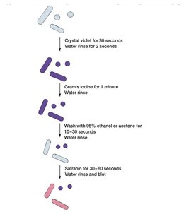
Gram Staining Steps:
Crystal violet (primary stain)
Iodine (mordant)
Alcohol or acetone (decolorizer)
Safranin (counterstain)
Result: Gram-positive cells remain purple; Gram-negative cells appear pink.
Special Stains
Special stains are used to visualize specific structures such as capsules, endospores, and flagella.
Capsule stain: Negative staining highlights the capsule as a clear halo around the cell.
Endospore stain: Endospores appear green within red or pink cells after staining with malachite green and safranin.
Flagella stain: Uses a mordant and dye to thicken flagella, making them visible under the microscope.
Example: Capsule staining is important for identifying pathogenic bacteria with protective capsules.
Summary Table: Types of Staining
Stain Type | Main Purpose | Example Organism/Structure |
|---|---|---|
Simple Stain | Visualize cell shape and arrangement | Any bacterial cell |
Gram Stain | Differentiates Gram+ and Gram- bacteria | Staphylococcus aureus (Gram+), Escherichia coli (Gram-) |
Acid-Fast Stain | Identifies acid-fast bacteria | Mycobacterium tuberculosis |
Capsule Stain | Visualizes bacterial capsules | Klebsiella pneumoniae |
Endospore Stain | Detects endospores | Bacillus species |
Flagella Stain | Visualizes flagella | Proteus species |
Additional info: Mastery of microscopy and staining techniques is foundational for identifying and studying microorganisms in clinical and research settings.
