Ch. 19 - Pathogenic Gram-Positive Bacteria
Bauman - Microbiology with Diseases by Taxonomy 6th Edition
Bauman6th EditionMicrobiology with Diseases by TaxonomyISBN: 9780134832302Non è quello che usi tu?Cambia libro di testo
Capitolo 19, Problema 6

Explain why Gram-positive mycoplasmas appear pink in a Gram-stained smear.

Guida verificata passo dopo passo
1
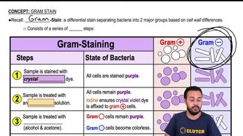
Recall that the Gram stain differentiates bacteria based on the structure of their cell walls: Gram-positive bacteria have a thick peptidoglycan layer that retains the crystal violet-iodine complex, appearing purple, while Gram-negative bacteria have a thinner peptidoglycan layer and do not retain the crystal violet, appearing pink after counterstaining with safranin.
Understand that mycoplasmas are unique bacteria that lack a cell wall entirely, meaning they do not have the peptidoglycan layer necessary to retain the crystal violet stain during the Gram staining process.
Since mycoplasmas lack the thick peptidoglycan layer, the crystal violet-iodine complex is washed away during the decolorization step of the Gram stain procedure.
After decolorization, mycoplasmas take up the counterstain safranin, which is pink/red, causing them to appear pink in the Gram-stained smear despite being classified as Gram-positive based on phylogeny.
Therefore, the pink appearance of Gram-positive mycoplasmas in a Gram stain is due to their lack of a cell wall, which prevents retention of the primary stain and results in uptake of the counterstain.

Risposta video verificata per un problema simile:

Questa soluzione video è stata consigliata dai nostri tutor come utile per risolvere questo problema.
Durata del video:
2m

Concetti chiave

Ecco i concetti essenziali che devi comprendere per rispondere correttamente alla domanda.

Gram Staining Mechanism

Gram staining differentiates bacteria based on cell wall structure. Gram-positive bacteria have thick peptidoglycan layers that retain the crystal violet-iodine complex, appearing purple, while Gram-negative bacteria have thinner walls and appear pink after counterstaining with safranin.
Video consigliato:
04:57
Gram Stain

Cell Wall Structure of Mycoplasmas

Mycoplasmas lack a cell wall entirely, including peptidoglycan, which is essential for retaining the crystal violet stain. This absence causes them to not retain the primary stain and instead take up the counterstain, appearing pink like Gram-negative bacteria.
Video consigliato:
02:59
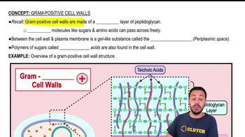
Gram-Positive Cell Walls

Limitations of Gram Staining for Mycoplasmas

Because mycoplasmas lack a cell wall, Gram staining is unreliable for their identification. Their pink appearance in Gram stains is due to the uptake of the counterstain, not their Gram reaction, highlighting the need for alternative diagnostic methods.
Video consigliato:
04:57
Gram Stain