Capillary permeability increase and plasma proteins leak into the interstitial fluid as part of the inflammatory process. Why is this desirable?
Ch. 11 Cardiovascular System

11장, 문제 22
How does the heart's ability to contract differ from that of other muscles of the body?
검증된 단계별 안내1
Understand the unique properties of cardiac muscle: Cardiac muscle is specialized for continuous, rhythmic contraction. Unlike skeletal muscle, which requires neural stimulation to contract, cardiac muscle has intrinsic contractility due to the presence of pacemaker cells in the sinoatrial (SA) node.
Learn about the role of pacemaker cells: The SA node generates electrical impulses automatically, without external stimulation. These impulses spread through the heart, causing coordinated contraction. This property is called autorhythmicity, which is unique to cardiac muscle.
Compare cardiac muscle to skeletal muscle: Skeletal muscle contraction is voluntary and requires signals from the nervous system. In contrast, cardiac muscle contraction is involuntary and self-regulated, though it can be influenced by the autonomic nervous system to adjust heart rate and force of contraction.
Explore the role of intercalated discs: Cardiac muscle cells are connected by intercalated discs, which contain gap junctions and desmosomes. Gap junctions allow electrical impulses to pass quickly between cells, ensuring synchronized contraction, while desmosomes provide structural support during contraction.
Understand the importance of calcium in cardiac contraction: Calcium plays a critical role in cardiac muscle contraction. The influx of calcium ions during an action potential triggers the release of more calcium from the sarcoplasmic reticulum, leading to contraction. This process is known as calcium-induced calcium release, which is distinct from the mechanism in skeletal muscle.

비슷한 문제에 대한 검증된 영상 답변:
이 영상 해법은 위 문제에 도움이 된다고 튜터들이 추천한 것입니다.
영상 길이:
2m도움이 되었나요?
주요 개념
질문에 올바르게 답하기 위해 반드시 이해해야 하는 핵심 개념들은 다음과 같습니다.
Cardiac Muscle Physiology
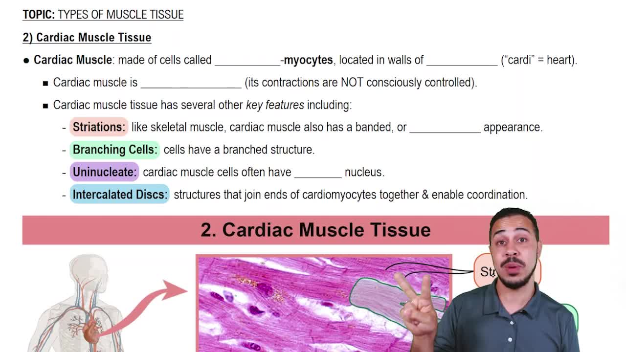
Cardiac muscle is unique in its structure and function, characterized by intercalated discs that allow for synchronized contractions. Unlike skeletal muscle, which is under voluntary control, cardiac muscle operates involuntarily and is influenced by the autonomic nervous system and hormones. This specialization enables the heart to pump blood efficiently and rhythmically.
추천 영상:
Cardiac Muscle Tissue
Excitation-Contraction Coupling
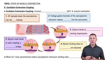
Excitation-contraction coupling refers to the physiological process by which an electrical stimulus leads to muscle contraction. In cardiac muscle, this process involves the influx of calcium ions through voltage-gated channels, triggering further calcium release from the sarcoplasmic reticulum. This mechanism is crucial for the heart's ability to contract and relax in a coordinated manner.
추천 영상:
B. Excitation-Contraction Coupling
Pacemaker Cells
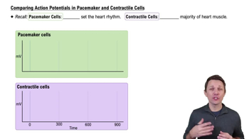
Pacemaker cells, primarily located in the sinoatrial (SA) node of the heart, are specialized cardiac cells that generate electrical impulses autonomously. These impulses initiate the contraction of the heart muscle, setting the rhythm for the heartbeat. This intrinsic ability distinguishes cardiac muscle from skeletal muscle, which requires neural stimulation for contraction.
추천 영상:
Comparing Action Potentials in Pacemaker and Contractile Cells
관련 실천
교과서 질문
1645
views
교과서 질문
Draw a diagram of the heart showing the three layers composing its wall and its four chambers. Label each. Show where the AV and semilunar valves are, and name them. Show and label all blood vessels entering and leaving the heart chambers.
1490
views
교과서 질문
Name three different factors that increase heart rate.
842
views
2
rank
교과서 질문
More than one choice may apply.
Using the figure, determine which vessels have the most resistance.
a. Arteries
b. Veins
c. Capillaries
d. Arterioles
e. Venules
898
views
교과서 질문
Name three factors that are important in promoting venous return.
166
views
교과서 질문
Trace one drop of blood from the time it enters the right atrium of the heart until it enters the left atrium. What is this circuit called?
1597
views