 Back
BackAcellular Pathogens: Viruses, Viroids, Virusoids, and Prions
Study Guide - Smart Notes
 Tailored notes based on your materials, expanded with key definitions, examples, and context.
Chapter 6: Acellular Pathogens
6.1 Viruses
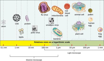
Viruses are acellular infectious agents that are fundamentally different from cellular life forms. They are obligate intracellular parasites, meaning they require a host cell to replicate. Viruses are extremely small, ranging from 20 nm to 900 nm, and are not visible with light microscopes.
Acellular: Viruses lack cellular structures such as ribosomes, mitochondria, cell membrane, cell wall, and chloroplasts.
Obligate Intracellular Parasites: Viruses can only reproduce inside living host cells.
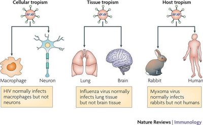
Host Range: Most viruses are host-specific, infecting only certain species and cell types.
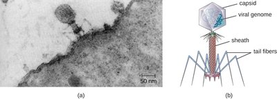
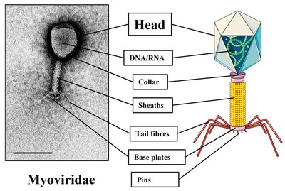
Bacteriophages: Viruses that infect bacteria are called bacteriophages or phages.
Effects on Host Cells: Viruses may cause abnormal growth, cell death, alter the cell’s genome, or cause little noticeable effect.
Hosts and Viral Transmission
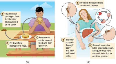
Viruses can infect a wide variety of hosts, including animals, plants, fungi, and bacteria. Transmission can occur through direct contact, indirect contact via fomites, or through vectors.
Zoonoses: Diseases transmitted from animals to humans.
Reverse Zoonoses: Diseases transmitted from humans to animals.
Fomites: Inanimate objects that can carry and spread infectious agents (e.g., doorknobs, towels, syringes).
Vectors: Animals (often arthropods) that transmit pathogens. Mechanical vectors carry pathogens on their body; biological vectors transmit pathogens through biting.


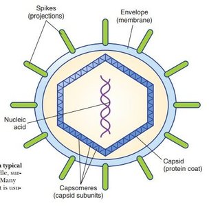
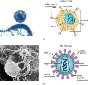
Viral Structures
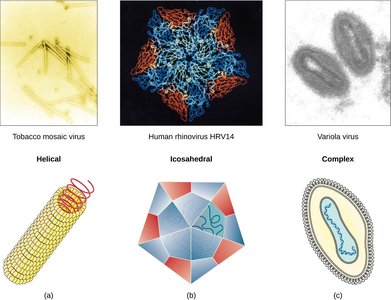
Viruses consist of genetic material (DNA or RNA, never both), a protein coat called a capsid, and sometimes a lipid envelope. The capsid is made of protein subunits called capsomeres. Some viruses have spikes (protein projections) for attachment to host cells.
Capsid Shapes: Helical, polyhedral (icosahedral), and complex.
Envelope: Some viruses have a phospholipid envelope derived from the host cell membrane.
Spikes: Protein structures for attachment and entry (e.g., hemagglutinin and neuraminidase in influenza viruses).
6.2 The Viral Life Cycle
Life Cycle of Viruses with Prokaryotic Hosts
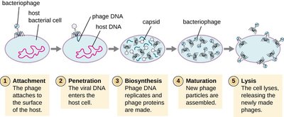
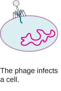
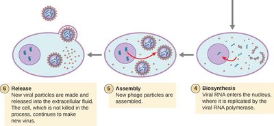
Bacteriophages replicate only in the cytoplasm of prokaryotic cells. There are two main types of phage life cycles: lytic and lysogenic.
Lytic Cycle: Virulent phages take over the cell, reproduce new phages, and destroy the cell via lysis.
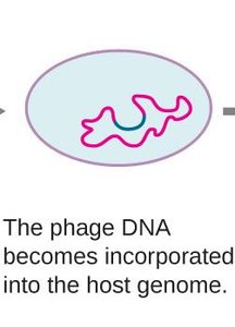
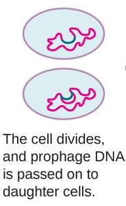
Lysogenic Cycle: Temperate phages integrate their genome into the host chromosome as a prophage, replicating with the cell until induced to enter the lytic cycle.
Transduction
Transduction is the process by which a bacteriophage transfers bacterial DNA from one bacterium to another during sequential infections. There are two types: generalized (random DNA transfer) and specialized (specific DNA transfer).

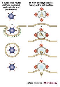
Life Cycle of Viruses with Animal Hosts
Animal viruses interact with specific host cell receptors and enter cells via endocytosis or membrane fusion. The viral genome is released (uncoating), followed by biosynthesis, maturation, and release (by budding or lysis).
Tissue Tropism: Most viruses infect specific cell types within tissues.
Animal Virus Genome Types
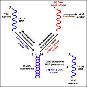
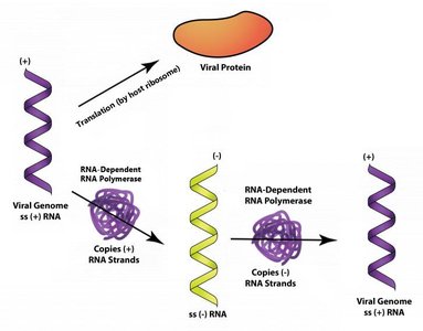
Animal viruses may have double-stranded DNA (dsDNA), single-stranded DNA (ssDNA), double-stranded RNA (dsRNA), or single-stranded RNA (ssRNA). ssRNA viruses can be positive-sense (+ssRNA) or negative-sense (-ssRNA).
+ssRNA: Can be directly translated by host ribosomes.
-ssRNA: Must be replicated into +ssRNA by viral RNA-dependent RNA polymerase before translation.
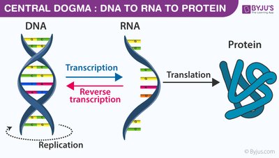
Retroviruses: +ssRNA viruses that use reverse transcriptase to synthesize DNA from RNA, integrating into the host genome as a provirus.
Persistent Infections
Persistent infections occur when a virus is not completely cleared from the host, remaining in tissues or organs. They are categorized as latent (dormant, asymptomatic) or chronic (recurrent, persistent symptoms).
Latent Viruses: Herpesviridae family (e.g., herpes simplex, varicella-zoster).
Chronic Viruses: Hepatitis C, HIV.
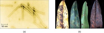
Life Cycle of Viruses with Plant Hosts
Plant viruses are similar to animal viruses and may be enveloped or non-enveloped, with DNA or RNA genomes. Transmission occurs via contact between plants or mechanical vectors such as fungi, nematodes, insects, or arthropods.
Viral Growth Curve
The viral growth curve consists of inoculation (attachment), eclipse (entry of genome), and burst (release of new virions). Burst size is the maximum number of virions produced per bacterium.
6.3 Isolation, Culture, and Identification of Viruses
Isolation of Viruses
Viruses require living host cells for replication. Infected host cells can be cultured and harvested as a source of virus. Virions can be separated from host cells by centrifugation or filtration.
Cultivation of Viruses
Viruses can be grown in vivo (within living organisms) or in vitro (in artificial environments such as cell cultures).
In Vivo: Diagnosis, vaccine production, and research using embryonated eggs or whole animals.
In Vitro: Primary cell cultures (limited lifespan) and continuous cell lines (immortal, e.g., HeLa cell line).
Detection of Viruses
Several methods are used to detect viruses:
Cytopathic Effects (CPEs): Observable cell abnormalities due to viral infection.
Hemagglutination Assay: Detects viral hemagglutinins that agglutinate red blood cells.
Nucleic Acid Amplification Tests (NAAT): Detect unique viral nucleic acid sequences (PCR for DNA, RT-PCR for RNA).
Enzyme Immunoassay (EIA): Uses antibodies to detect viral antigens.
6.4 Viroids, Virusoids, and Prions
Viroids
Viroids are short strands of circular RNA capable of self-replication. They cause diseases in plants, such as potato tuber spindle disease, tomato planta macho viroid, avocado sunblotch viroid, and peach latent mosaic viroid.
Virusoids
Virusoids are subviral particles described as non–self-replicating satellite RNAs. They require helper viruses for replication and belong to a larger group of infectious agents called satellite RNAs.
Prions
Prions are proteinaceous infectious particles discovered by Stanley Prusiner. They cause transmissible spongiform encephalopathies (TSE) in humans and animals, which are rare degenerative disorders affecting the brain and nervous system. Prions are extremely resistant to destruction and are transmitted via heredity, contact with contaminated tissue, or consumption of contaminated meat.
Type | Structure | Host | Replication |
|---|---|---|---|
Virus | DNA or RNA, capsid, sometimes envelope | Animals, plants, bacteria | Obligate intracellular |
Viroid | Circular RNA | Plants | Self-replicating |
Virusoid | Satellite RNA | Plants (with helper virus) | Requires helper virus |
Prion | Protein | Animals | Protein misfolding |
Additional info: This summary expands on brief slide points to provide full academic context, definitions, and examples for each acellular pathogen type, as well as their life cycles, transmission, and detection methods.
