 Back
BackAdaptive Immunity: B Lymphocytes and Antibodies
Study Guide - Smart Notes
 Tailored notes based on your materials, expanded with key definitions, examples, and context.
Adaptive Immunity
B Lymphocytes (B Cells) and Antibodies
Adaptive immunity is a highly specific defense mechanism that develops in response to exposure to antigens. B lymphocytes (B cells) are a central component of this system, primarily responsible for the production of antibodies. These cells are found mainly in the spleen, lymph nodes, and mucosa-associated lymphoid tissue (MALT), with a smaller proportion circulating in the blood.
Major Function: Secretion of antibodies that bind specifically to antigens.
Location: Spleen, lymph nodes, MALT, and blood.
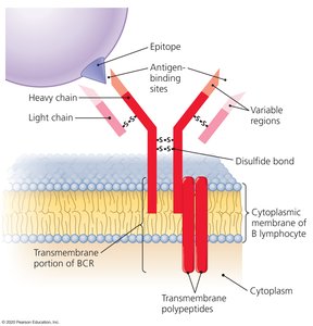
B Cell Receptor (BCR) Specificity
Each B lymphocyte expresses multiple copies of a unique B cell receptor (BCR) on its surface. The BCR is designed to recognize a specific epitope, which is a distinct part of an antigen. The diversity of BCRs in the body allows recognition of millions of different epitopes, but each individual B cell is specific for only one epitope.
Antigen-Binding Sites: Formed by two variable regions of the BCR.
Specificity: Each B cell recognizes only one epitope.
Diversity: The total BCR repertoire can recognize millions of epitopes.
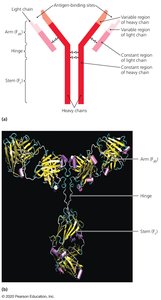
Antibody Structure and Secretion
Antibodies, also known as immunoglobulins, are secreted by activated B cells called plasma cells. The structure of an antibody is similar to that of the BCR, with antigen-binding sites and specificity identical to the BCR of the activated B cell.
Antigen-Binding Sites: Identical to those of the BCR from which the plasma cell originated.
Secretion: By plasma cells derived from B lymphocytes.
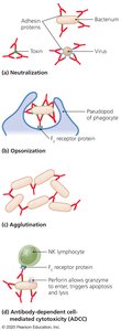
Antibody Functions
Antibodies perform several critical functions in the immune response by binding to antigens. Their antigen-binding sites are complementary to specific epitopes, enabling a range of immune activities:
Activation of Complement: Especially by IgM antibodies, leading to pathogen lysis.
Inflammation: IgE antibodies bind to mast cells/eosinophils, triggering release of inflammatory chemicals.
Neutralization: Antibodies block the activity of toxins or prevent pathogens from attaching to host cells.
Opsonization: IgG antibodies enhance phagocytosis by marking pathogens for ingestion.
Agglutination: Antibodies cause pathogens to clump together, facilitating removal.
Antibody-Dependent Cellular Cytotoxicity (ADCC): Antibodies recruit natural killer (NK) cells to destroy target cells.
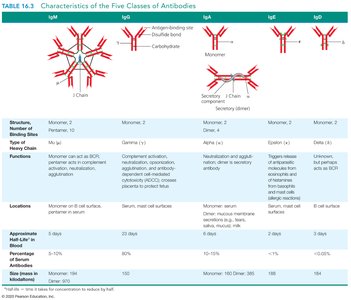
Classes of Antibodies
There are five main classes of antibodies, each with distinct roles in the immune response. The class involved depends on the type of antigen, the portal of entry, and the required immune function.
IgM: First antibody produced; effective in complement activation and agglutination.
IgG: Most common and longest-lasting antibody in the blood; provides the majority of antibody-based immunity.
IgA: Associated with mucosal surfaces and bodily secretions (e.g., saliva, tears, breast milk).
IgE: Involved in responses to parasitic infections and allergic reactions.
IgD: Function not fully understood; found on B cell surfaces.
Class | Structure | Functions | Locations |
|---|---|---|---|
IgM | Pentamer | Complement activation, agglutination, first antibody produced | Blood, B cell surface |
IgG | Monomer | Most abundant, crosses placenta, opsonization, neutralization | Blood, extracellular fluid |
IgA | Dimer (secretory) | Secretions, mucosal immunity | Secretions (saliva, tears, mucus) |
IgE | Monomer | Allergic reactions, defense against parasites | Bound to mast cells, basophils |
IgD | Monomer | Unclear, B cell receptor | B cell surface |
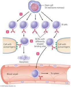
Clonal Deletion of B Cells
Clonal deletion is a process that ensures self-tolerance by eliminating or inactivating B cells that react against self-antigens. This process occurs in the bone marrow and is similar to the deletion of self-reactive T cells. Some self-reactive B cells may become inactive (anergic) or alter their BCRs instead of undergoing apoptosis.
Location: Bone marrow.
Outcome: Apoptosis, anergy, or receptor editing for self-reactive B cells.
Immune Response Cytokines
Cytokines are soluble regulatory proteins that serve as intercellular signals in the immune system. They are secreted by various leukocytes and form a complex network of communication among immune cells. Different types of cytokines have specialized roles:
Interleukins (ILs): Mediate communication between leukocytes.
Interferons (IFNs): Antiviral proteins that can also act as cytokines.
Growth Factors: Stimulate stem cell division and differentiation.
Tumor Necrosis Factor (TNF): Regulates immune responses, inflammation, and can induce apoptosis in tumor cells.
Chemokines: Direct the movement of immune cells (chemotaxis).
Representative Cytokines and Their Actions
Cytokine | Source | Target | Action |
|---|---|---|---|
Interleukin 2 (IL-2) | Th1 cell, Tc cell | Tc cell | Cloning of Tc cell |
Interleukin 4 (IL-4) | Th2 cell | B cell | B cell differentiates into plasma cell |
Interleukin 12 (IL-12) | Dendritic cell | Th cell | Th cell differentiates into Th1 cell |
Gamma interferon (IFN-γ) | Th1 cell | Macrophage | Increases phagocytosis |
Tumor necrosis factor (TNF) | Macrophages, T cells | Body tissues | Triggers inflammation or apoptosis |
Key Concepts Review
The antibody-binding site of an antibody is made up of: The variable regions of both the heavy and light chains.
The most prevalent antibody class in the blood is: IgG.
A single B lymphocyte can recognize multiple antigenic determinants: False. Each B lymphocyte recognizes only one antigenic determinant (epitope).
