 Back
BackAdaptive Immunity: Specific Defenses of the Host
Study Guide - Smart Notes
 Tailored notes based on your materials, expanded with key definitions, examples, and context.
Adaptive Immunity: Specific Defenses of the Host
Introduction to Adaptive Immunity
Adaptive immunity is a specialized defense mechanism that targets specific pathogens. Unlike innate immunity, which provides general protection, adaptive immunity develops after exposure to antigens through infection or vaccination. It is characterized by specificity and memory, allowing for a more rapid and effective response upon subsequent exposures to the same pathogen.
Primary response: The initial adaptive immune reaction to a novel antigen.
Secondary response: A faster, stronger reaction upon re-exposure to the same antigen, due to immunological memory.
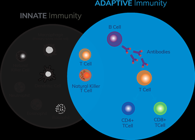
Dual Nature of the Adaptive Immune System
The adaptive immune system consists of two main components: humoral immunity and cellular immunity. Both systems interact with the innate immune system to provide comprehensive protection.
Humoral immunity: Mediated by B cells, which produce antibodies that neutralize extracellular pathogens and toxins.
Cellular immunity: Mediated by T cells, which recognize and destroy infected or abnormal cells.
Humoral Immunity
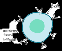
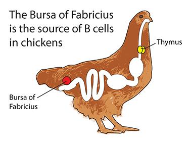
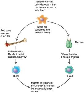
B cells: Lymphocytes that mature in the bone marrow and are responsible for antibody production.
Antibodies: Proteins that specifically bind to antigens, marking them for destruction.
Example: Neutralization of bacterial toxins by antibodies.
Cellular Immunity
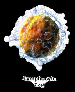
T cells: Lymphocytes that mature in the thymus and are involved in recognizing antigenic peptides presented by other cells.
T cell receptors (TCRs): Surface molecules that bind to specific antigens.
Cytokines: Secreted by T cells to direct immune responses.
Example: Destruction of virus-infected cells by cytotoxic T lymphocytes (CTLs).
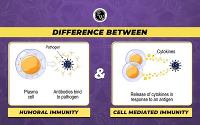
Comparison of Humoral and Cellular Immunity
Humoral immunity: Targets extracellular pathogens (e.g., bacteria, toxins, viruses before cell entry).
Cellular immunity: Targets intracellular pathogens (e.g., viruses, some bacteria inside host cells).
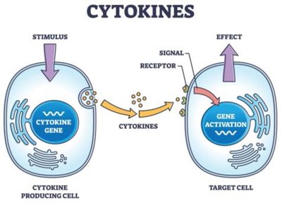
Cytokines: Chemical Messengers of Immune Cells
Cytokines are signaling proteins produced by immune cells in response to stimuli. They regulate the intensity and duration of immune responses by mediating communication between cells.
Interleukins (ILs): Mediate communication between leukocytes.
Chemokines: Induce migration of immune cells to infection sites.
Interferons (IFNs): Inhibit viral replication and activate immune cells.
Tumor necrosis factor alpha (TNF-α): Promotes inflammation, especially in autoimmune diseases.
Hematopoietic cytokines: Stimulate production of blood cells from stem cells.
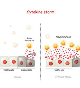
Cytokine storm: Overproduction of cytokines, leading to tissue damage.
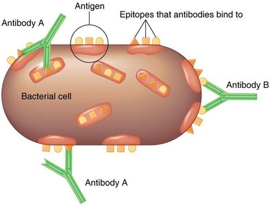
Antigens and Antibodies
Antigens are substances that provoke an immune response, typically by stimulating the production of antibodies. Antibodies recognize specific regions on antigens called epitopes.
Epitope: The specific part of an antigen recognized by an antibody.
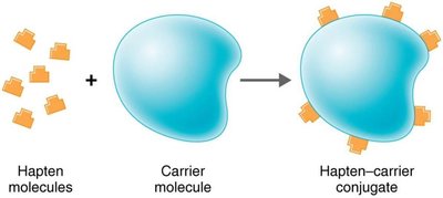
Hapten: A small molecule that is not immunogenic by itself but can elicit an immune response when attached to a carrier protein.
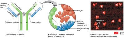
Structure and Classes of Antibodies (Immunoglobulins)
Antibodies are Y-shaped proteins composed of two heavy and two light chains, with variable regions that bind antigens and a constant region that determines the antibody class.
Valence: Number of antigen-binding sites (most antibodies are bivalent).
Variable (V) region: Binds to the epitope of the antigen.
Constant (Fc) region: Determines the antibody class and mediates effector functions.
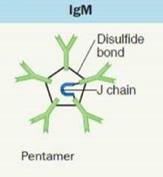
Five classes of immunoglobulins: IgG, IgM, IgA, IgD, IgE.

Summary Table: Immunoglobulin Classes
Class | Structure | Serum % | Main Functions |
|---|---|---|---|
IgG | Monomer | 80% | Crosses placenta, enhances phagocytosis, neutralizes toxins/viruses, activates complement |
IgM | Pentamer | 6% | First antibody produced, agglutinates microbes, activates complement |
IgA | Monomer/dimer | 13% | Protects mucosal surfaces, found in secretions (tears, saliva, breast milk) |
IgD | Monomer | 0.02% | On B cells, assists in immune response |
IgE | Monomer | 0.002% | Allergic reactions, defense against parasitic worms |

Humoral Immunity Response Process
B cells are activated to produce antibodies through interactions with antigens and helper T cells. This process involves antigen presentation, clonal selection, and differentiation into plasma and memory cells.
Major histocompatibility complex (MHC): Glycoproteins on cell surfaces that present antigen fragments to T cells.
Class I MHC: Present on all nucleated cells; recognized by CD8+ T cells.
Class II MHC: Present on antigen-presenting cells (APCs) such as B cells, macrophages, and dendritic cells; recognized by CD4+ T cells.
Clonal selection: Activated B cells proliferate and differentiate into antibody-producing plasma cells and memory cells.
Clonal deletion: Elimination of self-reactive B cells to prevent autoimmunity (via apoptosis).
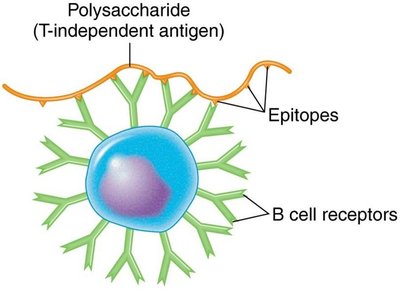
T-Dependent and T-Independent Antigens
T-dependent antigens: Require helper T cells for B cell activation; generate strong, long-lasting immunity with memory cell formation.
T-independent antigens: Activate B cells without T cell help; usually polysaccharides; provoke a weaker response, mainly IgM, and do not generate memory cells.
Results of Antigen-Antibody Interaction
When antibodies bind to antigens, several protective mechanisms are triggered to eliminate the pathogen:
Agglutination: Clumping of antigens, enhancing phagocytosis.
Opsonization: Coating of antigens to enhance phagocytosis.
Neutralization: Blocking of pathogen attachment or toxin activity.
Antibody-dependent cell-mediated cytotoxicity (ADCC): Destruction of large pathogens by immune cells.
Activation of the complement system: Leads to cell lysis.
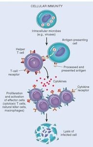
Cellular Immunity Response Process
Cellular immunity is mediated by T cells, which recognize and respond to intracellular pathogens. T cells mature in the thymus and are classified based on their surface markers and functions.
CD4+ T cells (Helper T cells): Activate B cells, other T cells, and macrophages via cytokine secretion.
CD8+ T cells (Cytotoxic T lymphocytes, CTLs): Destroy infected or abnormal cells by inducing apoptosis.
T regulatory cells (Treg): Suppress immune responses to maintain self-tolerance and prevent autoimmunity.
Subsets of Helper T Cells
TH1 cells: Activate macrophages and promote cell-mediated immunity.
TH2 cells: Stimulate B cells to produce IgE and activate eosinophils.
TH17 cells: Recruit neutrophils and promote inflammation.
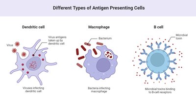
Antigen-Presenting Cells (APCs)
Dendritic cells: Capture and present antigens to T cells; found in tissues and blood.
Macrophages: Engulf pathogens and present antigens to T cells; activated by cytokines.
B cells: Present antigens to helper T cells for further activation.
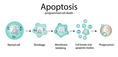
Apoptosis
Definition: Programmed cell death, a mechanism to eliminate infected or harmful cells without causing inflammation.
Nonspecific Cells and Extracellular Killing
Natural killer (NK) cells: Destroy cells lacking MHC class I molecules, such as virus-infected or tumor cells, by inducing lysis or apoptosis. NK cells are not antigen-specific.
Antibody-dependent cell-mediated cytotoxicity (ADCC): Immune cells recognize and kill antibody-coated target cells, important for defense against large parasites.
Immunological Memory
Immunological memory is the ability of the adaptive immune system to respond more rapidly and effectively to pathogens that have been encountered previously.
Primary response: Initial exposure to an antigen; slower and less robust.
Secondary (anamnestic) response: Subsequent exposure; faster, stronger, and longer-lasting due to memory cells.
Antibody titer: Measurement of antibody concentration in serum, reflecting the strength of the humoral response.
Types of Adaptive Immunity
Adaptive immunity can be acquired naturally or artificially, and can be active or passive:
Type | How Acquired | Example |
|---|---|---|
Naturally acquired active | Infection | Recovery from measles |
Naturally acquired passive | Maternal antibodies | Antibodies via placenta or breast milk |
Artificially acquired active | Vaccination | MMR vaccine |
Artificially acquired passive | Injection of antibodies | Gamma globulin injection |
