 Back
BackAdaptive Immunity: Specific Defenses of the Host
Study Guide - Smart Notes
 Tailored notes based on your materials, expanded with key definitions, examples, and context.
Adaptive Immunity: Specific Defenses of the Host
Introduction to Adaptive Immunity
Adaptive immunity is a specialized defense mechanism that targets specific pathogens. Unlike innate immunity, which provides general protection, adaptive immunity develops after exposure to antigens through infection or vaccination. It is characterized by specificity and memory, allowing for a more rapid and effective response upon subsequent exposures to the same pathogen.
Primary response: The initial adaptive immune reaction to a new antigen.
Secondary response: A faster, stronger reaction upon re-exposure to the same antigen, due to immunological memory.
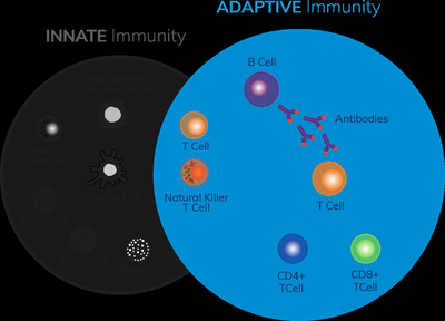
Comparison of Adaptive and Innate Immunity
Innate immunity: Non-specific, immediate defense present from birth (e.g., skin, phagocytes).
Adaptive immunity: Specific, acquired defense that develops after exposure to antigens; involves lymphocytes (B and T cells) and antibodies.
Acquisition of Adaptive Immunity
Immunity can be acquired naturally (through infection) or artificially (through vaccination).
Vaccination is an example of adaptive immunity, as it induces a specific immune response and memory formation.
Dual Nature of the Adaptive Immune System
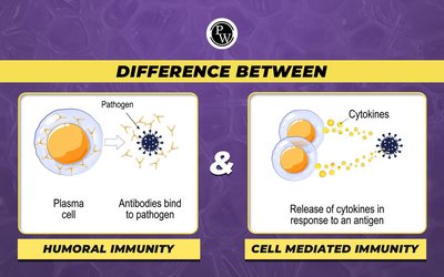
Humoral Immunity
Humoral immunity is mediated by B lymphocytes (B cells) and the antibodies they produce. It is primarily effective against extracellular pathogens and toxins.
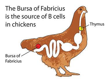
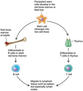
B cells: Lymphocytes that mature in the bone marrow and produce antibodies in response to antigens.
Antibodies: Proteins that specifically bind to antigens, neutralizing them or marking them for destruction.
Named for: The bursa of Fabricius in birds, where B cells were first identified.
Cellular Immunity (Cell-Mediated Immunity)
Cellular immunity is mediated by T lymphocytes (T cells), which mature in the thymus. T cells recognize and respond to antigens presented by infected or abnormal cells, primarily targeting intracellular pathogens such as viruses and some bacteria.
T cells: Lymphocytes that recognize antigenic peptides presented by major histocompatibility complex (MHC) molecules on other cells.
T cell receptors (TCRs): Surface molecules on T cells that bind to specific antigen-MHC complexes.
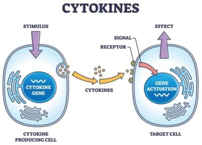
Cytokines: Chemical messengers secreted by T cells to direct immune responses.
Comparison of Humoral and Cellular Immunity
Humoral immunity: Fights invaders outside cells (e.g., extracellular bacteria, toxins, viruses before cell entry).
Cellular immunity: Targets pathogens inside cells (e.g., viruses, intracellular bacteria like Mycobacterium leprae).
Cytokines: Chemical Messengers of Immune Cells
Types and Functions of Cytokines
Cytokines are signaling proteins produced by immune cells in response to stimuli. They coordinate the activities of the immune system by mediating communication between cells.
Interleukins (ILs): Mediate communication between leukocytes.
Chemokines: Induce migration of leukocytes to infection sites.
Interferons (IFNs): Interfere with viral replication in host cells.
Tumor necrosis factor alpha (TNF-α): Involved in inflammation and autoimmune diseases.
Hematopoietic cytokines: Regulate development of blood cells from stem cells.
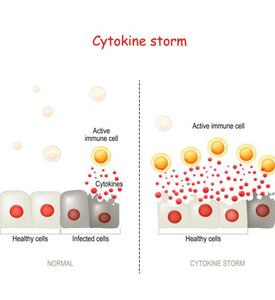
Cytokine storm: Overproduction of cytokines can lead to tissue damage and severe inflammation.
Antigens and Antibodies
Antigens, Epitopes, and Haptens
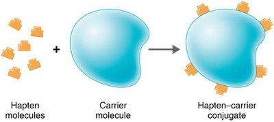
Antigens are substances that provoke an immune response, typically by stimulating the production of antibodies. They are usually components of pathogens or foreign substances. Epitopes are specific regions on antigens that antibodies recognize and bind to. Haptens are small molecules that are not immunogenic by themselves but can elicit an immune response when attached to a larger carrier molecule.
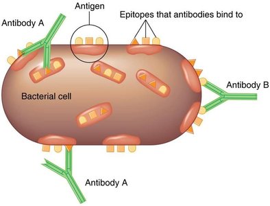
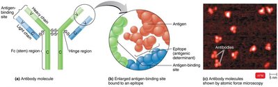
Structure and Function of Antibodies
Antibodies, or immunoglobulins (Ig), are Y-shaped proteins composed of two identical heavy chains and two identical light chains, joined by disulfide bonds. Each antibody has two antigen-binding sites (valence), allowing it to bind specifically to epitopes on antigens. The variable (V) regions at the tips of the Y arms determine antigen specificity, while the constant (Fc) region determines the antibody class and mediates effector functions.
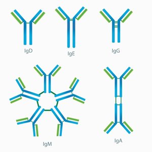
Classes of Immunoglobulins
Class | Structure | Serum % | Main Functions |
|---|---|---|---|
IgG | Monomer | 80% | Crosses placenta, protects fetus, triggers complement, enhances phagocytosis, neutralizes toxins/viruses |
IgM | Pentamer | 6% | First antibody produced, causes agglutination, activates complement |
IgA | Monomer/dimer | 13% | Found in mucous membranes, saliva, tears, breast milk; prevents microbial attachment |
IgD | Monomer | 0.02% | On B cells; assists in immune response |
IgE | Monomer | 0.002% | On mast cells/basophils; involved in allergic reactions and defense against parasites |
Humoral Immunity Response Process
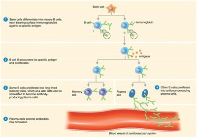
Activation of B Cells and Antibody Production
B cells are activated when their surface immunoglobulins bind to specific antigens. The antigen is internalized, processed, and presented on MHC class II molecules. Helper T cells (TH) recognize the antigen-MHC complex and secrete cytokines that stimulate B cell proliferation (clonal expansion) and differentiation into plasma cells (which secrete antibodies) and memory cells (which provide long-term immunity).
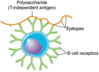
T-Dependent and T-Independent Antigens
T-dependent antigens: Require help from TH cells to activate B cells; generate strong, long-lasting immune responses and memory cells.
T-independent antigens: Can activate B cells without TH cell help; usually polysaccharides; provoke weaker responses and do not generate memory cells.
Clonal Selection and Deletion
Clonal selection: Process by which activated B cells proliferate and differentiate into plasma cells and memory cells.
Clonal deletion: Elimination of self-reactive B cells to prevent autoimmune reactions (via apoptosis).
Results of Antigen-Antibody Interaction
When antibodies bind to antigens, several protective mechanisms are triggered:
Agglutination: Clumping of antigens, enhancing phagocytosis.
Opsonization: Coating of antigens to enhance phagocytosis.
Neutralization: Blocking of toxins or viruses from binding to host cells.
Activation of complement system: Leads to cell lysis.
Antibody-dependent cell-mediated cytotoxicity (ADCC): Target cell is lysed by immune cells.
Cellular Immunity Response Process
T Cell Maturation and Function
T cells mature in the thymus, where thymic selection eliminates self-reactive cells. Mature T cells migrate to lymphoid tissues and recognize antigens presented by MHC molecules on antigen-presenting cells (APCs).
CD4+ T cells (Helper T cells): Recognize antigens presented by MHC class II; secrete cytokines to activate other immune cells.
CD8+ T cells (Cytotoxic T lymphocytes, CTLs): Recognize antigens presented by MHC class I; kill infected or abnormal cells by inducing apoptosis.
T Helper Cell Subsets
TH1 cells: Activate macrophages and CTLs; promote cell-mediated immunity.
TH2 cells: Stimulate B cells to produce IgE; activate eosinophils.
TH17 cells: Recruit neutrophils; involved in inflammation.
T regulatory cells (Treg): Suppress immune responses to maintain self-tolerance and prevent autoimmunity.
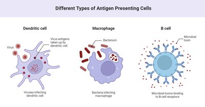
Antigen-Presenting Cells (APCs)
Dendritic cells: Engulf and present antigens to T cells; found in tissues and blood.
Macrophages: Present antigens after phagocytosis; activated by cytokines.
B cells: Can also act as APCs.
Apoptosis
Programmed cell death (apoptosis) is a mechanism by which CTLs and NK cells eliminate infected or abnormal cells, preventing the spread of infection.
Nonspecific Cells and Extracellular Killing
Natural Killer (NK) Cells
NK cells are granular leukocytes that destroy cells lacking MHC class I self-antigens. They kill virus-infected and tumor cells, and attack parasites. NK cells can induce apoptosis or lysis in target cells, and participate in antibody-dependent cell-mediated cytotoxicity (ADCC).
Immunological Memory
Primary and Secondary Immune Responses
Primary response: Occurs after first exposure to an antigen; slower and less intense.
Secondary (anamnestic) response: Occurs upon re-exposure; faster, stronger, and longer-lasting due to memory cells.
Class switching: Initial IgM response shifts to IgG, IgE, or IgA during secondary response.
Antibody titer: Measurement of antibody concentration in serum, reflecting the intensity of the humoral response.
Types of Adaptive Immunity
Type | How Acquired | Example |
|---|---|---|
Naturally acquired active | Infection | Recovery from measles |
Naturally acquired passive | Maternal antibodies (placenta, breast milk) | IgG transfer to fetus |
Artificially acquired active | Vaccination | MMR vaccine |
Artificially acquired passive | Injection of antibodies | Gamma globulin injection |
