Back
BackAdaptive Immunity: Structure, Function, and Mechanisms
Study Guide - Smart Notes
 Tailored notes based on your materials, expanded with key definitions, examples, and context.
Adaptive Immunity
Overview and Comparison with Innate Immunity
Adaptive immunity is the body's highly specific defense mechanism against distinct invaders and their products. It is distinguished from innate immunity by its specificity, inducibility, clonality, unresponsiveness to self, and memory. The following table compares innate and adaptive immunity:
Feature | Innate Immunity | Adaptive Immunity |
|---|---|---|
Distribution | Almost all multicellular eukaryotes | Only in vertebrates |
Targets | Limited number of key structures (PAMPs) | Antigens (billions of types) |
Immune Receptors | Pattern recognition receptors (e.g., TLRs) | T cell receptors and antibodies |
Cellular Presence | Almost all cells | Lymphocytes only |
Discrimination | Host cells lack PAMPs | Tolerance for self-antigens can break down (autoimmunity) |
Immunological Memory | Absent | Present |
Key Attributes of Adaptive Immunity
Specificity: Targets unique antigens.
Inducibility: Activated only in response to specific pathogens.
Clonality: Generates clones of lymphocytes specific to the antigen.
Unresponsiveness to Self: Prevents attack on self-antigens.
Memory: Remembers previous encounters for faster response.
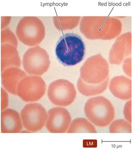
Lymphocytes in Adaptive Immunity
Lymphocytes are central to adaptive immunity. There are two main types:
B lymphocytes (B cells): Mature in the bone marrow; responsible for antibody production.
T lymphocytes (T cells): Mature in the thymus; responsible for cell-mediated responses.
Adaptive immunity involves two main responses:
Cell-mediated immune responses: Target intracellular pathogens and abnormal cells.
Antibody (humoral) immune responses: Target exogenous pathogens and toxins.
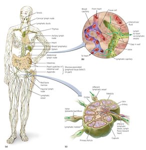
The Lymphatic System
Tissues and Organs of the Lymphatic System
The lymphatic system screens the tissues of the body for foreign molecules and is composed of lymphatic vessels, lymphoid cells, tissues, and organs.
Lymphatic vessels: One-way system conducting lymph from tissues back to the circulatory system.
Lymph: Fluid similar to blood plasma, arising from leaked blood vessel fluid.
Primary lymphoid organs: Red bone marrow and thymus.
Secondary lymphoid organs: Lymph nodes, spleen, tonsils, and mucosa-associated lymphoid tissue (MALT).
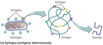
Antigens and Their Properties
Definition and Structure
Antigens are molecules recognized as foreign and worthy of attack. They are identified by three-dimensional regions called epitopes.
Best antigens: Large foreign macromolecules.
Sources: Bacterial components, proteins of viruses, fungi, protozoa, food, and dust.
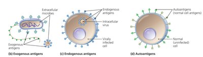
Types of Antigens
Exogenous antigens: Toxins and components of microbial cell walls, membranes, flagella, and pili.
Endogenous antigens: Produced by microbes reproducing inside body cells.
Autoantigens: Derived from normal cellular processes.
Major Histocompatibility Complex (MHC) and Antigen Presentation
MHC Proteins
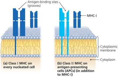
MHC proteins are glycoproteins found in the membranes of most vertebrate cells. They hold and position antigenic epitopes for presentation to immune cells.
MHC class I: Present on all cells except red blood cells.
MHC class II: Present on antigen-presenting cells (APCs) such as macrophages, B cells, and dendritic cells.
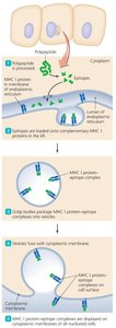
Antigen Processing
Antigens are processed for MHC proteins to display epitopes. The process differs for endogenous and exogenous antigens.
T Lymphocytes (T Cells)
Production, Maturation, and Migration
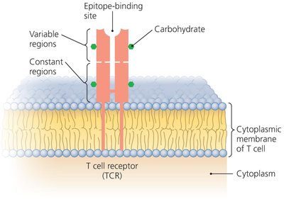
T cells are produced in the red bone marrow, mature in the thymus, and migrate to lymph nodes, spleen, and Peyer's patches. They possess T cell receptors (TCRs) on their membrane.
Specificity of TCR
TCRs bind only epitopes associated with MHC proteins.
T cells act primarily against cells harboring intracellular pathogens or abnormal cell-surface proteins.
Types of T Lymphocytes
Cytotoxic T lymphocyte (Tc): Directly kills other cells.
Helper T lymphocyte (Th): Regulates B cells and cytotoxic T cells; includes Th1 and Th2.
Regulatory T lymphocyte (Tr): Represses adaptive immune responses.
Lymphocyte | Site of Maturation | Representative Cell-Surface Glycoproteins | Notable Secretions |
|---|---|---|---|
Helper T cell type 1 (Th1) | Thymus | CD4, distinctive TCR | Interleukin 2, IFN-γ |
Helper T cell type 2 (Th2) | Thymus | CD4, distinctive TCR | Interleukin 4, 5 |
Cytotoxic T cell (Tc) | Thymus | CD8, CD95L, distinctive TCR | Perforin, granzyme |
Regulatory T cell (Tr) | Thymus | CD4, CD25, distinctive TCR | Interleukin 10 |
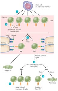
Clonal Deletion of T Cells
Clonal deletion is vital to prevent immune responses against autoantigens. Self-reactive lymphocytes are eliminated by apoptosis.
T cells not recognizing MHC protein undergo apoptosis.
T cells recognizing autoantigen die by apoptosis.
Some self-recognizing T cells become regulatory T cells.
T cells recognizing MHC protein and foreign epitopes become protective T cells.
B Lymphocytes (B Cells) and Antibodies
Production and Function
B cells are found primarily in the spleen, lymph nodes, and MALT. Their major function is the secretion of antibodies.
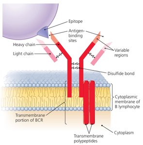
Specificity of B Cell Receptor (BCR)
Each B cell generates a single BCR.
Two variable regions form antigen-binding sites.
Each BCR recognizes only one epitope.
The repertoire of BCRs can recognize millions of different epitopes.
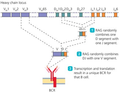
Lymphocyte Receptor Diversity
Genes for constant and variable regions of BCRs occur at discrete loci. The RAG enzyme combines different variable region gene segments to generate antibody diversity.
Antibody Structure and Function
Antibodies are immunoglobulins similar to BCRs, secreted by plasma cells. They have antigen-binding sites and specificity identical to the BCR of the activated B cell.
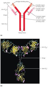
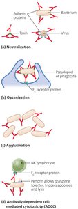
Functions: Activation of complement and inflammation, neutralization, opsonization, agglutination, antibody-dependent cellular cytotoxicity (ADCC).
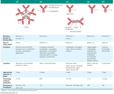
Classes of Antibodies
IgM: First antibody produced.
IgG: Most common and longest-lasting antibody.
IgA: Associated with body secretions.
IgE: Involved in response to parasitic infections and allergies.
IgD: Exact function unknown.
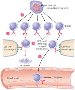
Clonal Deletion of B Cells
Clonal deletion occurs in the bone marrow. Self-reactive B cells may become inactive or change their BCR rather than undergo apoptosis.
Immune Response Cytokines
Cytokine Types and Functions
Cytokines are soluble regulatory proteins acting as intercellular signals. They are secreted by various leukocytes and form a complex network of signals.
Interleukins (ILs): Signal among leukocytes.
Interferons (IFNs): Antiviral proteins, may act as cytokines.
Growth factors: Stimulate stem cells to divide.
Tumor necrosis factor (TNF): Kills tumor cells, regulates immune responses and inflammation.
Chemokines: Signal leukocytes to move.
Cytokine | Source | Target | Action |
|---|---|---|---|
Interleukin 2 (IL-2) | Th1 cell, Tc cell | Tc cell | Cloning of Tc cell |
Interleukin 4 (IL-4) | Th2 cell | B cell | B cell differentiates into plasma cell |
Interleukin 12 (IL-12) | Dendritic cell | Th cell | Th cell differentiates into Th1 cell |
Gamma interferon (IFN-γ) | Th1 cell | Macrophage | Increases phagocytosis |
Tumor necrosis factor (TNF) | Macrophages, T cells | Body tissues | Triggers inflammation or apoptosis |
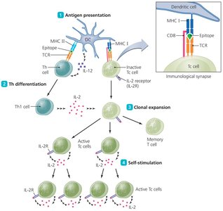
Cell-Mediated Immune Responses
Activation of Cytotoxic T Cell Clones
Cell-mediated responses target intracellular pathogens and abnormal body cells. Activation of cytotoxic T cells involves four steps:
Antigen presentation
Helper T cell differentiation
Clonal expansion
Self-stimulation
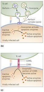
Mechanisms of Cytotoxic T Cell Killing
Perforin-granzyme pathway: Synthesis of killing proteins.
CD95 pathway: Mediated through glycoprotein on body’s cells.
Memory T Cells
Some activated T cells become memory T cells, persisting for months or years in lymphoid tissues. They respond immediately upon subsequent contact with the specific epitope-MHC complex.
T Cell Regulation
Regulation prevents T cell response to autoantigens. T cells require additional signals from antigen-presenting cells, and regulatory T cells moderate cytotoxic T cell activity.
Antibody Immune Responses
T-Dependent Antibody Immunity and Clonal Selection
T-dependent antibody immunity depends on helper T cells and involves four steps:
Antigen presentation for Th activation and proliferation
Differentiation of helper T cells into Th2 cells
Activation of B cells
Proliferation and differentiation of B cells
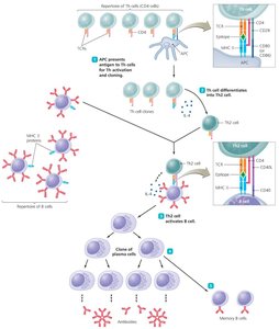
Plasma Cells and Memory Cells
Plasma cells: Secrete antibody molecules complementary to the specific antigen; short-lived but their antibodies and progeny persist.
Memory B cells: Produced by B cell proliferation; do not secrete antibodies but initiate antibody production if antigen is encountered again.
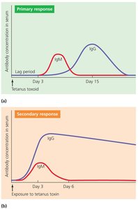
Primary and Secondary Immune Responses
Primary response: Small amounts of antibodies produced; may take days to eliminate antigen.
Secondary response: Memory cells respond rapidly to another exposure; much faster than primary response.
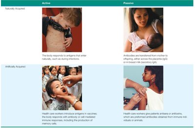
Types of Acquired Immunity
Active vs. Passive Immunity
Specific immunity is acquired during an individual's life and can be:
Naturally acquired: Response against antigens encountered in daily life.
Artificially acquired: Response to antigens introduced via a vaccine.
Active: Body produces its own antibodies.
Passive: Antibodies are received from another source.