 Back
BIO 120 Exam I Study Guide: Microbiology Essentials
Study Guide - Smart Notes
 Tailored notes based on your materials, expanded with key definitions, examples, and context.
Introductory Concepts
Microbiology as a Science
Microbiology is the study of microscopic organisms, including bacteria, viruses, fungi, and protozoa. It explores their structure, function, and role in health and disease.
Pasteurization: A process developed by Louis Pasteur to kill harmful microbes in food and drinks, especially milk, by heating them to a specific temperature.
Germ Theory of Disease: Proposes that microorganisms are the cause of many diseases. Robert Koch's four postulates are used to establish a causal relationship between a microbe and a disease.
Koch's Postulates:
The microorganism must be found in all organisms suffering from the disease, but not in healthy organisms.
The microorganism must be isolated from a diseased organism and grown in pure culture.
The cultured microorganism should cause disease when introduced into a healthy organism.
The microorganism must be re-isolated from the inoculated, diseased experimental host and identified as being identical to the original specific causative agent.
Microscopy
Microscopy is essential for visualizing microorganisms. Different types of microscopes offer varying resolution and magnification.
Resolution vs Magnification: Resolution is the ability to distinguish two points as separate; magnification is the enlargement of an image.
Bright Field: Standard light microscope; best for stained specimens.
Dark Field: Enhances contrast in unstained samples; useful for live organisms.
Phase Contrast: Highlights differences in refractive index; ideal for live cells.
Fluorescence: Uses fluorescent dyes; allows visualization of specific structures.
SEM (Scanning Electron Microscope): Provides 3D images of surfaces.
TEM (Transmission Electron Microscope): Offers detailed internal structure images.
Stains
Types of Stains and Their Importance
Staining techniques are used to enhance contrast and differentiate between types of microorganisms.
Simple Stain: Uses a single dye to highlight cells.
Differential Stains:
Gram Stain: Differentiates bacteria into Gram-positive (purple) and Gram-negative (pink) based on cell wall structure.
Acid Fast Stain: Identifies mycobacteria, such as Mycobacterium tuberculosis.
Special Stains:
Endospore Stain: Detects bacterial endospores.
Capsule Stain: Negative stain highlights capsules.
Flagellar Stain: Visualizes bacterial flagella.
Bacterial Morphology and Arrangement
Shapes and Groupings
Bacteria are classified by their shape and arrangement.
Bacillus: Rod-shaped; can be diplo- (pairs) or strepto- (chains).
Coccus: Spherical; can be diplo-, staphylo- (clusters), or strepto- (chains).
Spiral: Includes vibrio (comma-shaped), spirillum (rigid spiral), and spirochete (flexible spiral).
Prokaryotic Structures
Cell Components and Functions
Prokaryotes have unique structures that support their survival and function.
Membrane: Selectively permeable; site of energy production.
Chromosome: Free-floating in cytoplasm; plasmids are extra-chromosomal DNA.
Fimbriae and Pili: Used for attachment, motility, and genetic transfer.
Cytoplasm: Contains cellular components.
Flagella: Responsible for motility; arrangement can be peritrichous, amphitrichous, lophotrichous, or polar.
Cell Wall: Provides protection, rigidity, and shape; Gram-positive vs Gram-negative based on peptidoglycan structure.
Glycocalyx: Includes capsule, slime layer, and biofilms.
Endospores: Highly resistant dormant structures.
Ribosomes: Protein synthesis; structurally different from eukaryotic ribosomes.
Inclusion Bodies: Storage of nutrients and other substances.
Metachromatic granules: Phosphate reserves
Polysaccharide granules: Energy reserves
Lipid inclusions: Energy reserves
Sulfur granules: Energy reserves
Carboxysomes: CO2 fixation
Gas vacuoles: Buoyancy
Magnetosomes: Iron oxide, destroys H2O2
Eukaryotic Structures
Cell Components and Functions
Eukaryotic cells have complex organelles and structures.
Membrane: Selectively permeable.
Cell Wall: Composed of cellulose or chitin; absent in animal cells.
Cytoplasm: Contains cytoskeleton (microtubules and actin) for movement.
Nucleus: Membrane-bound; contains DNA and nuclear pores for RNA export.
Endoplasmic Reticulum (ER): Smooth ER (lipid synthesis), Rough ER (protein synthesis).
Golgi Apparatus: Modifies proteins and lipids for transport.
Plasma Membrane: Contains integral, transmembrane, and peripheral proteins.
Flagella: Membrane-bound; 9+2 microtubule arrangement.
Cilia: Similar to flagella; used for movement and mucus removal.
Endosymbiotic Theory: Organelles evolved from symbiosis between prokaryotes and primitive eukaryotes. Evidence includes similarities between mitochondria/chloroplasts and bacteria.
Viruses
Structure and Life Cycles
Viruses are acellular entities that require a host for replication. They can use DNA or RNA as genetic material and may be enveloped or non-enveloped.
Virion vs Virus: A virion is the complete, infectious form of a virus outside a host cell.
Enveloped vs Non-enveloped: Enveloped viruses enter cells via membrane fusion; non-enveloped viruses use receptor-mediated endocytosis.
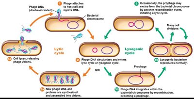
Bacteriophage: Viruses that infect bacteria; exhibit lytic and lysogenic cycles.
Lytic Cycle: Virus replicates and lyses host cell, releasing new virions.
Lysogenic Cycle: Viral DNA integrates into host genome, replicates with cell, and can later enter lytic cycle.
Animal Viruses: Includes HIV and retroviruses; targets for drug intervention.
Antiviral Drugs vs Antibiotics: Antivirals target viral replication; antibiotics target bacteria.
Latency: Viruses can remain dormant (e.g., herpes viruses).
Oncoviruses: Can cause cancer by disrupting cell cycle regulation genes.
Protozoa and Fungi
Protozoa
Protozoa are single-celled eukaryotes with diverse modes of movement and disease causation.
Movement:
Ciliates: Move by cilia
Amebae: Move by pseudopodia
Flagellates: Move by flagella
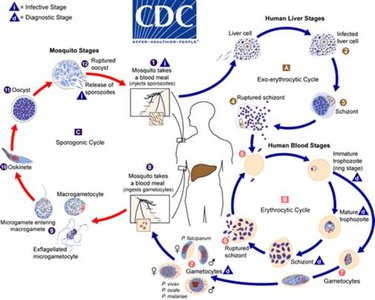
Diseases: Examples include malaria and giardiasis.
Fungi
Fungi are eukaryotic organisms that include yeasts, molds, and fleshy fungi.
Yeasts: Divide by budding.
Molds: Have aerial and vegetative hyphae; dimorphic fungi can be yeast or hyphae.
Fungal Reproduction: Can be sexual or asexual.
Diseases
Types of Fungal Diseases
Fungal diseases are classified by the site of infection.
Cutaneous: Affect skin (e.g., athlete's foot, ringworm).
Subcutaneous: Affect deeper skin layers (e.g., Madura foot).
Systemic: Affect internal organs.
Respiratory: Example: Histoplasma capsulatum.
