 Back
BackCardiovascular & Lymphatic Infections: Systemic Infections in Microbiology
Study Guide - Smart Notes
 Tailored notes based on your materials, expanded with key definitions, examples, and context.
Overview of the Cardiovascular & Lymphatic Systems
Basic Structure and Function
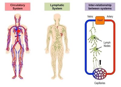
The cardiovascular system and lymphatic system are interconnected networks responsible for transporting blood, lymph, nutrients, immune factors, and waste products throughout the body. These systems play a critical role in both health and disease, particularly in the dissemination of infectious agents.
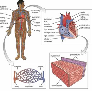
Cardiovascular System: A closed network of the heart and blood vessels that circulates blood to deliver oxygen, nutrients, and immune cells, and to remove waste products.
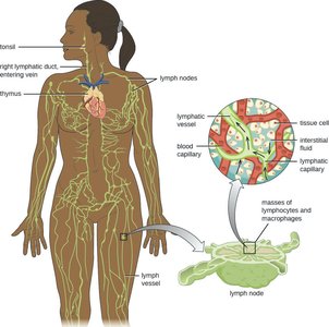
Lymphatic System: An open, one-way system that drains excess fluid (lymph) from tissues, filters it through lymph nodes, and returns it to the venous blood supply. It is rich in immune cells such as B-cells, T-cells, and macrophages.
Systemic Infections: Infections of these systems can rapidly become systemic, spreading microorganisms throughout the body.
Blood, Plasma, and Lymph
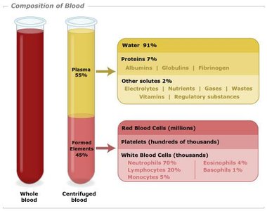
Blood: Composed of plasma, red blood cells (RBCs), white blood cells (WBCs), and platelets. It delivers oxygen, nutrients, and immune factors, and removes waste.
Plasma: The liquid portion of blood, containing proteins, hormones, nutrients, and dissolved gases.
Lymph: Plasma that has diffused into tissues, mixed with WBCs, and is collected by lymphatic vessels for immune surveillance and return to circulation.
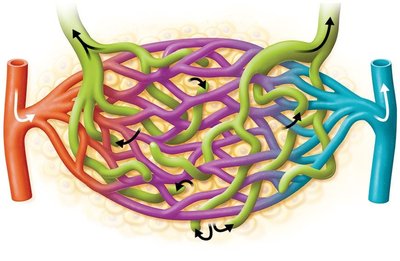
Lymphatic System Details
Open system, not pumped like the cardiovascular system; fluid moves in one direction toward veins above the heart.
Lymph nodes filter lymph and are sites of immune activation. Severe infection/inflammation of nodes is called bubos.
Edema: Swelling due to lymph accumulation in tissues.
Systemic Infections and Pathways
Microorganisms can spread systemically via the cardiovascular and lymphatic systems, either from localized infections (e.g., lung, kidney, GI, skin) or direct introduction (e.g., wounds, catheters, insect bites).
Key Definitions
Bacteremia/Viremia/Parasitemia: Presence of bacteria/viruses/parasites in the blood.
Septicemia: Microbes present and multiplying in the blood.
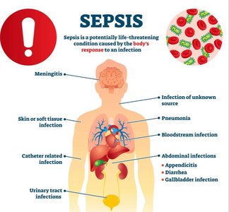
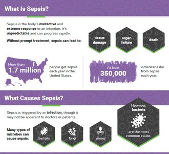
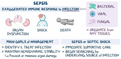
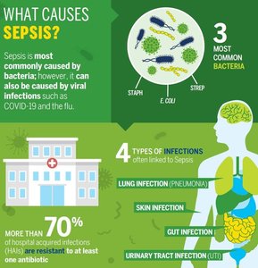
Sepsis: Life-threatening organ dysfunction caused by a dysregulated host response to infection or toxins, often leading to tissue and organ damage.
Septic Shock: Severe sepsis with dangerously low blood pressure, risking multiorgan failure and death.
Sepsis
Overview and Pathogenesis
Sepsis is not an infection itself but a severe, life-threatening condition resulting from the body's overwhelming and dysregulated immune response to infection or microbial toxins. It can progress to septic shock and multiorgan failure.
Common risk factors: Immunocompromised state, recent surgery, invasive procedures, severe trauma.
Not transmissible; prevention relies on effective infection management.
Signs and symptoms: Diarrhea, vomiting, pale skin, sleepiness, confusion, decreased urine output, delirium.
Diagnosis: Sequential Organ Failure Assessment (SOFA) score ≥2; septic shock includes severe hypotension.
Treatment: Reduce inflammation, stabilize blood pressure, increase oxygenation, administer antimicrobials if nonviral infection suspected.
Systemic Viral Infections
Arboviruses: Focus on Zika Virus
Arboviruses are arthropod-borne viruses, primarily transmitted by mosquitoes such as Aedes aegypti. Major arboviruses include dengue, yellow fever, chikungunya, and Zika.
Warming climates expand mosquito habitats, increasing arbovirus transmission risk.
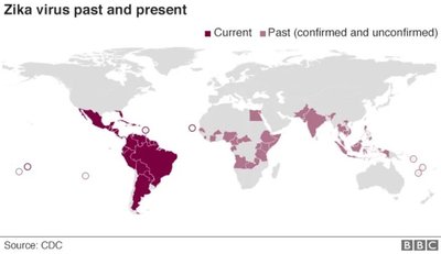
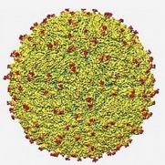
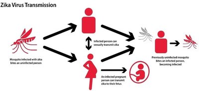
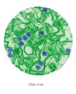
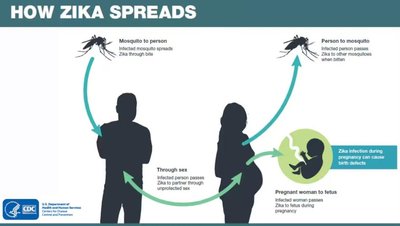
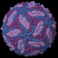
Zika Virus: Enveloped, single-stranded RNA virus (Flaviviridae). First identified in 1947; first U.S. cases in 2016.
Transmission: Mosquito bites (main route), sexual contact, blood transfusion, vertical (mother to fetus).
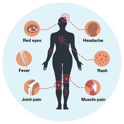
Symptoms: Often asymptomatic; when present, fever, joint pain, red eyes, rash.
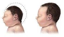
Complications: Vertical transmission can cause microcephaly and brain defects in fetuses.
Diagnosis: Serologic or molecular tests (blood, urine, saliva, semen).
Treatment: Supportive only; prevention includes mosquito control and delaying conception after infection (8 weeks for women, 6 months for men).
Hemorrhagic Viruses: Focus on Ebola
Hemorrhagic viruses cause severe fevers and bleeding by interfering with blood-clotting mechanisms. Notable examples include Lassa, Marburg, and Ebola viruses.
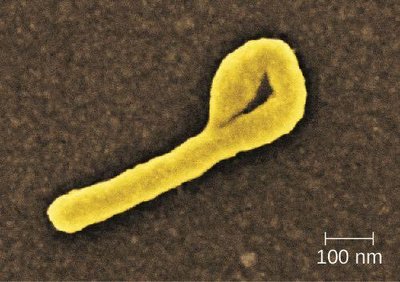
Ebola Virus: Linear, filamentous, enveloped, single-stranded RNA virus (Filoviridae). Highly contagious and lethal.
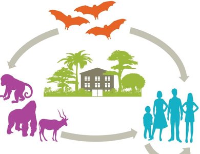
Transmission: Zoonotic (fruit bats as reservoir), contact with infected animals or bushmeat, person-to-person via bodily fluids, contaminated fomites.
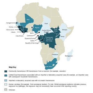
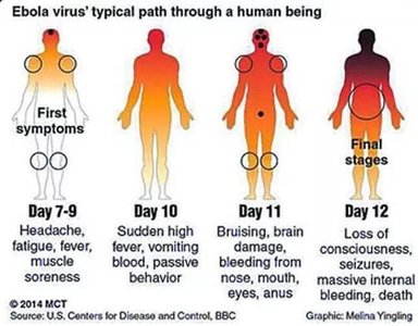
Epidemiology: Mainly in Central and West Africa; long incubation (2–21 days); highly transmissible and fatal.
Symptoms: Initial "dry phase" (fever, weakness, sore throat), followed by "wet phase" (hemorrhagic symptoms, organ failure, shock, death).
Treatment: Supportive therapy (rehydration, pain relief); vaccine (Ervebo) available for Ebola Zaire strain.
Prevention: Avoid contact with infected individuals/animals, use of PPE, vaccination in outbreak areas.
Retroviruses: Focus on HIV
Retroviruses are RNA viruses that use reverse transcriptase to integrate their genome into host DNA, forming a provirus. Human retroviruses include HIV and HTLV-1.
HIV: Enveloped RNA virus, targets CD4+ T helper cells, causes chronic infection and immunodeficiency (AIDS).
Transmission: Sexual contact, blood, IV drug use, vertical (mother to child).
Stages of Infection:
Stage 1 (Acute): Flu-like symptoms, high viral load, detectable by antigen/NAAT tests.
Stage 2 (Chronic): Clinically asymptomatic, slow viral replication, gradual CD4+ decline.
Stage 3 (AIDS): CD4+ count <200 cells/μl, high viral load, opportunistic infections/cancers.
Treatment: Combination antiretroviral therapy (ART) with at least three drugs; lifelong therapy required due to provirus persistence.
Prevention: Safe sex, needle hygiene, pre- and post-exposure prophylaxis (PrEP, PEP), no effective vaccine yet.
Systemic Bacterial Infections
Yersinia pestis: Plague
Yersinia pestis: Gram-negative bacillus, zoonotic (reservoir: rats, vector: fleas).
Three clinical forms:
Bubonic plague: Infects lymphatic system, causes bubos.
Septicemic plague: Bacteria in bloodstream, can cause sepsis, gangrene.
Pneumonic plague: Infects lungs, only form transmissible person-to-person, nearly 100% fatal if untreated.
Transmission: Flea bites (primary), direct contact with infected animals, inhalation of droplets.
Diagnosis: Laboratory/molecular testing of clinical specimens.
Treatment: Rapid antibiotic therapy; untreated plague is often fatal.
Prevention: Rodent control, flea prevention, prophylactic antibiotics for exposed individuals.
Borrelia burgdorferi: Lyme Disease
Borrelia burgdorferi: Gram-negative spirochete, transmitted by Ixodes (deer) ticks.
Three stages:
Early localized: Erythema migrans (bull's-eye rash), flu-like symptoms.
Early disseminated: Fatigue, additional rashes, cardiac and neurologic symptoms.
Late persistent: Severe arthritis, chronic neurologic and cardiac dysfunction.
Diagnosis: Clinical signs (rash, facial palsy, arthritis), history of tick exposure, lab tests (serology, NAATs).
Treatment: Antibiotics (2–4 weeks) curative in early stages; late-stage damage may be permanent.
Prevention: Avoid tick habitats, use repellents, perform tick checks, prompt tick removal, single-dose antibiotics after high-risk exposure.
Systemic Protozoan Infections
Plasmodium falciparum: Malaria
Malaria: Life-threatening disease caused by Plasmodium species, transmitted by Anopheles mosquitoes.
P. falciparum: Most common and lethal species, widespread in Africa and Asia.
Transmission: Mosquito bite injects sporozoites, which infect liver and then red blood cells (RBCs).
Symptoms: Cyclic fever, chills, headache, body aches, nausea, vomiting; severe cases include anemia, hypotension, hypoglycemia, acidosis, organ failure, coma.
Diagnosis: Microscopic blood analysis, rapid antigen tests.
Treatment: Antimalarial drugs (choice depends on species, region, resistance patterns); early treatment reduces mortality and transmission.
Prevention: Mosquito avoidance (repellents, nets, protective clothing), chemoprophylaxis, vaccination (Mosquirix for children in endemic areas).
Stage | Key Features |
|---|---|
Uncomplicated Malaria | Cycles of fever, chills, sweating (6–10 hours), repeat every 2–3 days |
Complicated Malaria | Anemia, hypotension, hypoglycemia, acidosis, organ failure, coma |
